Transesophageal echocardiography (TEE) is an advanced imaging technique that uses ultrasound waves via a probe inserted through the esophagus to visualize the heart closely. It provides clearer images than transthoracic echocardiography in specific clinical conditions.
TEE is commonly used in detecting heart valve diseases, congenital cardiac anomalies, and atrial thrombi. It is also essential in the evaluation of prosthetic valves and monitoring patients during cardiac surgeries.
In suspected endocarditis, TEE offers high diagnostic accuracy by detecting vegetations and abscesses that may not be visible on standard echocardiography. This makes it a gold standard method in certain infectious cardiac conditions.
While generally safe, TEE may cause temporary throat discomfort or minor esophageal irritation. In rare cases, complications like bleeding or perforation may occur, thus requiring careful application in selected patients.
| Medical Name | Transesophageal Echocardiography (TEE) |
| Common Uses | – Detailed evaluation of valvular heart diseases – Investigation for intracardiac clots, infections or tumors – Diagnosis of congenital heart diseases |
| Causes | – Inadequate visualization by transthoracic echocardiography – Suspected valvular heart disease – Suspected intracardiac infection (endocarditis) |
| Risk Factors | – Swallowing difficulties – Esophageal diseases (pharyngeal hernia, tumor, severe reflux, etc.) – Bleeding disorders |
| Complications | – Irritation or pain in the throat – Very rarely esophageal injury – Nausea, vomiting – Bleeding (rare) |
| Diagnostic Methods | – Preliminary evaluation with transthoracic echocardiography – Clinical examination |
| Treatment Methods | – Light sedation and local anesthesia during the procedure – Monitoring and expert supervision during the procedure |
| Prevention Methods | – Pre-procedure fasting (at least 6 hours) – Careful evaluation in patients with swallowing difficulties or esophageal disease |
What is TEE in Medicine? How Does This Device Show the Interior of the Heart?
The principle of TEE is actually quite simple: it uses the power of sound waves. At its center is a thin, flexible tube (probe) with a tiny ultrasound camera at the end. This probe sends out high-frequency sound waves that the human ear cannot hear. These sound waves hit your heart and the structures inside it (valves, chambers, blood cells) and are reflected back. The probe collects these returned “echoes” and sends them to the sophisticated computer system to which it is connected. The computer combines these echoes to create a vivid, moving and detailed image of your heart on the screen.
The real power of this technology is the ability to look at the heart through different “lenses”. Each mode allows us to see a different feature of the heart. The different imaging modes offered by TEE are usually the following:
- 2-Dimensional (2D) Imaging
- M-Mode Imaging
- Color Doppler
- Pulse Wave (PW) Doppler
- Continuous Wave (CW) Doppler
- 3-Dimensional (3D) Imaging
2D imaging shows snapshots of your heart, while Doppler modes present the flow, speed and direction of your blood through the heart in color, like a traffic map. 3D imaging takes it a step further, allowing surgeons to have a three-dimensional, beating model of your heart at their fingertips before surgery. This is an invaluable guide, especially for complex cover repairs.
How is it different from a normal chest echo? Why sometimes the answer to the question ‘How to do TEE’ becomes more important?
The natural curiosity of patients is why, when there is a standard echocardiogram performed through the chest, there is a need for a different method, TEE. The answer lies in the huge difference in image quality and the special areas TEE reaches. Normal ECHO has to send sound waves through the chest wall. But on this journey, the sound waves encounter many obstacles. Lungs, ribs and, especially in overweight individuals, subcutaneous fatty tissue act as barriers for sound waves. This can result in blurred images or some areas of the heart not being visible at all. We call this a “bad acoustic window”, like trying to look out of a dirty window.
TEE removes all of these obstacles. Because the probe is placed directly behind the heart through the esophagus, there are no obstacles in between. This results in much clearer and higher resolution images. This clarity makes a critical difference in diagnosing some vital conditions. The following are situations in which TEE is far superior to normal echo:
- Intra-heart clot detection
- Heart valve infections (Endocarditis)
- Prosthetic valve problems
- Aortic vessel ruptures
- Congenital heart holes
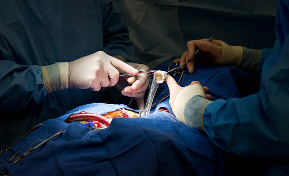
- Guidance during heart surgery
In such cases, the answer to the question “How is TEE performed?” becomes vital for proper diagnosis and treatment, as normal ECHO is inadequate. In these cases, TEE is not only a diagnostic tool but also a compass that determines the direction of treatment.
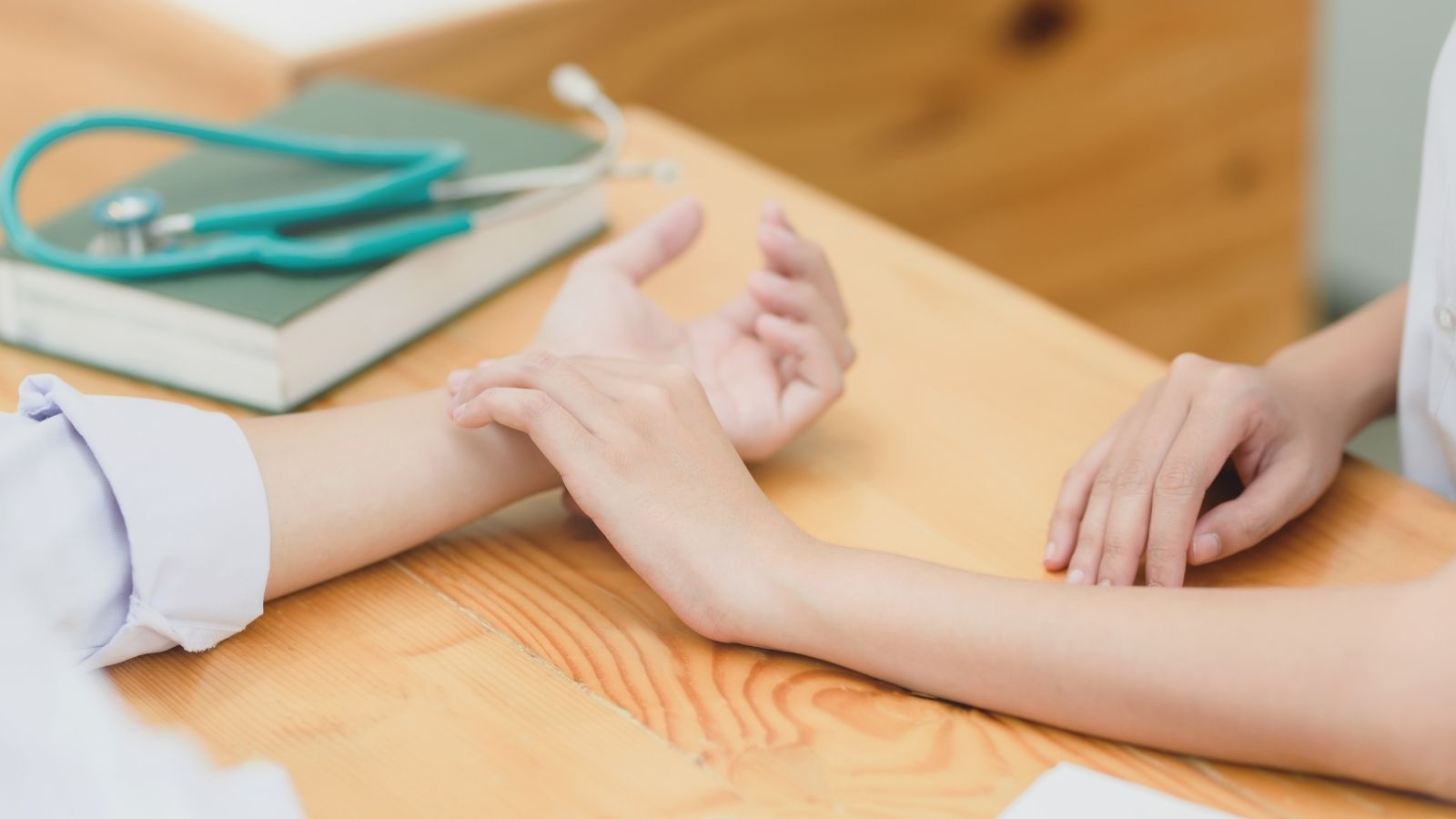
How is the Curious TEE Test Performed and What Does the Preparation Process Involve?
The most important question on many people’s minds is how this procedure is performed. The process is carefully planned, prioritizing patient comfort and safety. Knowing what to expect on the day of the procedure and beforehand will reduce your worries.
Pre-Process Preparation Phase
You will be asked to make some preparations to ensure that your transaction goes smoothly and safely. Preparations to be made before the procedure:
- 6-8 hours solid food hunger
- 2-4 hours fluid hunger
- Notification of medicines used
- Reporting of existing diseases (especially swallowing/stomach)
- Removal of dentures
- Signing the procedure consent form
The most important of these preparations is hunger. An empty stomach eliminates the risk of nausea and vomiting during the procedure and prevents the contents from entering your lungs (aspiration). This is a standard protocol for your security.
Process Day
Once you are in the procedure room, the process usually proceeds in the following steps. First, a mild sedative is given through a vein in your arm to help you relax and make the procedure comfortable. This medicine will not put you completely to sleep, it will only give you a pleasant drowsiness and a feeling of relaxation. Most patients do not remember clearly after the procedure. A numbing spray is then sprayed into your throat to suppress your gag reflex and ease the passage of the probe. This is an important step that can taste a bit bitter but makes the job much easier. A plastic mouthguard is placed between your teeth to protect both your teeth and the sensitive probe of the device.
When the doctor asks you to swallow, he or she gently guides the tip of the probe into your esophagus. This is the most critical moment in the whole process and is done with great precision, using absolutely no force. Once the probe is in the right place, imaging begins. The procedure usually takes between 15 and 30 minutes, during which time your heart rhythm, blood pressure and oxygen levels are monitored intermittently.
Is he completely asleep during the procedure? Like the video of TEE on the heart in the operating room?
This is an important question that depends on where and why TEE is performed. The images you can see on the internet, especially those posted under the title “video of TEE on the heart” are usually taken during cardiac surgery, which is completely different from the diagnostic TEE performed in the outpatient clinic.
Diagnostic TEE: If your doctor orders TEE to diagnose a disease, it is usually done in an endoscopy unit or echocardiography laboratory. In this case, general anesthesia is not used. Instead, a method called “conscious sedation” is used. Thanks to the intravenous medication, you will feel less anxious, relaxed and slightly drowsy. You may hear conversations around you, but they will not disturb you. When the procedure is over, you usually have little or no recollection of what happened. The goal is to keep your safety at the highest level while ensuring your comfort.
TEE During Heart Surgery: TEE used during heart surgery is a completely different scenario. The patient is already under general anesthesia for the surgery and is completely asleep. The TEE probe is inserted after the patient is asleep and on a ventilator. It guides the surgeon through the surgery and the patient feels absolutely nothing during this process. Therefore, the situation in those videos is a procedure applied only to patients under general anesthesia.
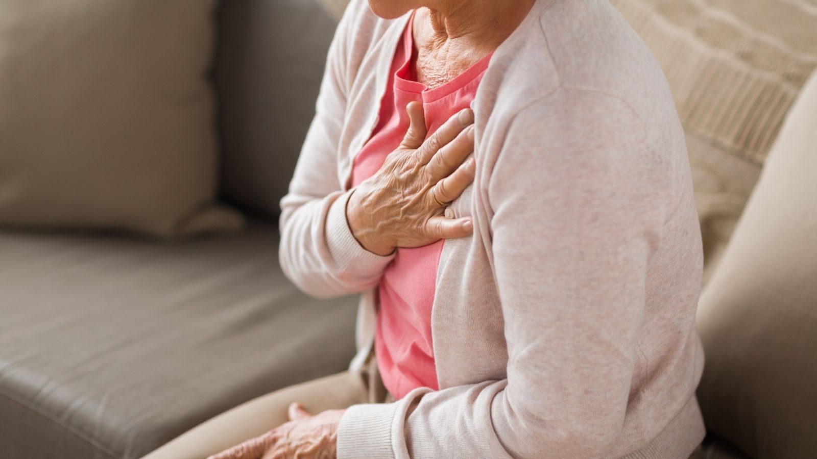
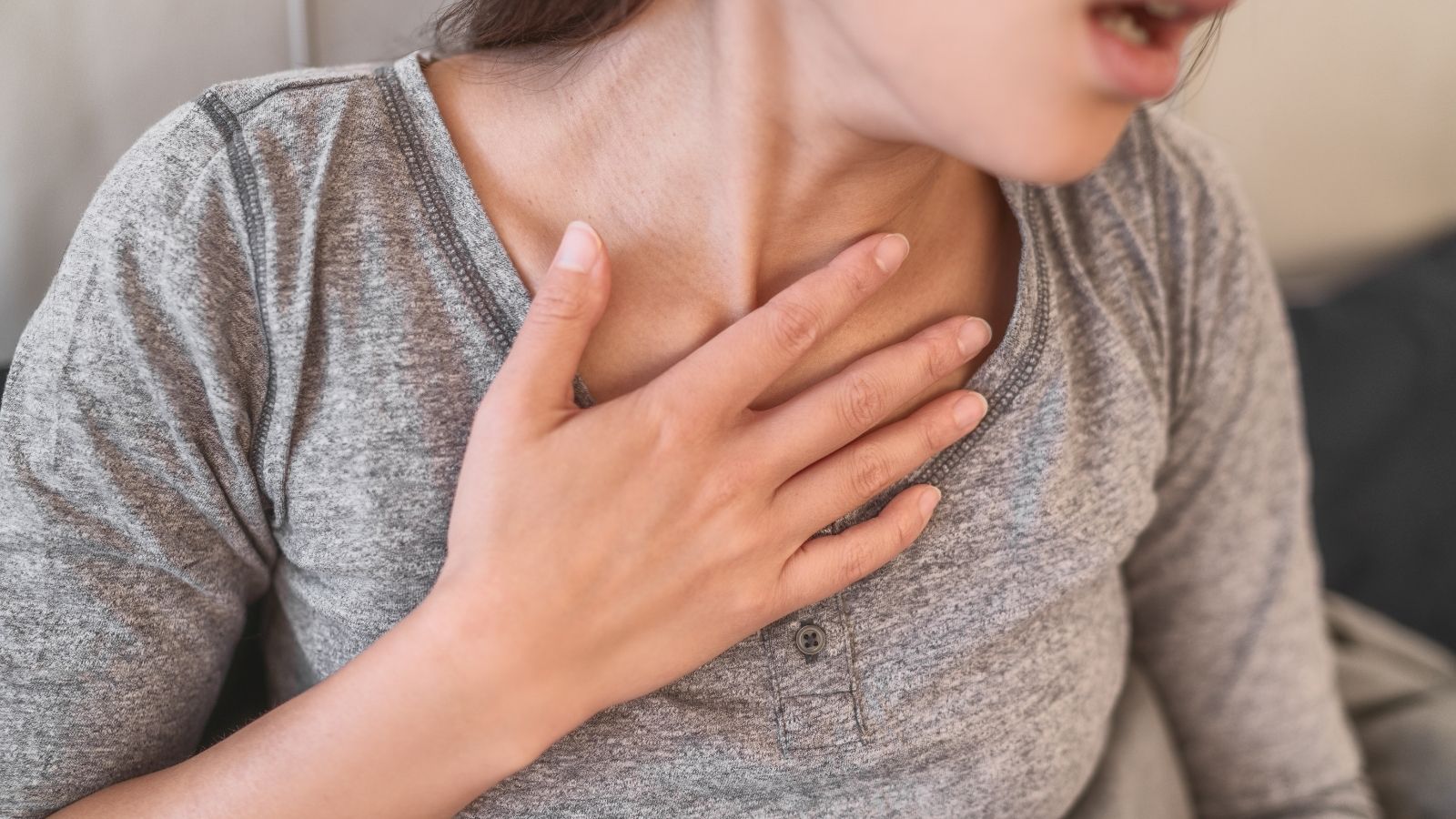
Should it be feared? What are the Side Effects of the TEE Test?
It is normal to have some anxiety before any medical procedure. being transparent about “TEE test side effects” is the best way to alleviate these concerns. First of all, it is important to emphasize the following: TEE is an extremely safe procedure when performed in experienced hands and with the right patient selection. The risk of serious complications is quite high.
The possible effects of the procedure can be divided into two groups. The most common side effects are usually mild:
- Pain or tenderness in the throat
- Temporary swallowing difficulty
These symptoms usually go away on their own within a day or two and do not cause any major discomfort. There are also risks that are much rarer but need to be recognized:
- Damage to the teeth
- Low blood pressure due to drugs
- Respiratory slowdown due to drugs
- Stricture or bleeding in the esophagus
- Perforation of the esophagus (very rare)
Perforation of the esophagus, the most serious of these risks, is more likely, especially in people with pre-existing esophageal disease. This is why a detailed consultation with you before the procedure is vital. Your doctor and his team will take all precautions to minimize these risks and will follow you closely throughout the procedure. Remember that the vital information TEE can provide often far outweighs these very low risks.
In Which Heart Diseases Does TEE Diagnosis Save Lives?
The unparalleled clarity provided by TEE makes it a key player in the diagnosis and follow-up of some complex and dangerous heart diseases.
Infective Endocarditis (Heart Valve Infection)
In this disease, clumps of germs and clots (vegetations) form on the heart valves. TEE is much more sensitive than normal echo in detecting these clumps. More importantly, it shows whether the infection has spread to the tissues around the valve. Dangerous conditions that TEE reveals in this disease:
- Vegetation (Germ culture)
- Abscess (pus accumulation)
- Fistula (abnormal connection)
- Cover puncture
- Prosthetic valve separation
Prosthetic Lid Problems
Prosthetic heart valves can create a “shadow” on normal echo due to their metallic structure, making a clear assessment difficult. TEE transcends this shadow and provides a clear view behind and around the prosthesis. Problems detected with TEE in prosthetic valves:
- Clot formation
- Pannus (Tissue growth)
- Cover edge paper (Paravalvular leak)
- Infection
Stroke and Clot Source Research
In patients who have had a stroke of unknown cause, TEE is the most reliable method to investigate whether the clot originates in the heart. Clot sources sought by TEE in stroke patients:
- Left atrial appendage (LAA) clot
- Patent foramen ovale (PFO)
- Plaques in the aorta
- Intracardiac tumors (rare)
TEE best visualizes the blind sac called the left atrial appendage (LAA) where most of the clots form, especially in patients with atrial fibrillation arrhythmias. Making sure there are no clots in this area is essential for many treatments to be carried out safely.
What are the comments of those who have had a TEE test and what do they mean?
Before a procedure, it is very human to be curious about what others who have had the experience have to say. when examining “comments from those who have had a TEE test”, certain themes often emerge. These comments both give an idea to the people who will undergo the procedure and point out the important points of the process.
Generally, the comments of those who have performed a TEE test center around the following issues:
- The process is easier than expected
- Comfort thanks to calming medication
- Mild throat pain after the procedure
- Clarity and relief at the result
The most important message to take away from these comments is this: The comfort and success of the procedure is highly dependent on the sedation and the experience of the team. The fact that patients describe the process as “easier than expected” is usually the result of a procedure administered by a professional team with the correct dosage of sedatives. Side effects such as sore throat are usually temporary and tolerable. Most importantly, patients often feel a sense of relief that an uncertain condition has been clarified and a diagnosis made. These comments suggest that TEE is not a procedure to be feared, but rather an important and bearable step to be taken for accurate diagnosis.
What Are TEE Test Prices Based on in 2025 and What Do They Depend on?
The cost of a TEE procedure is another topic that patients and their relatives are rightly curious about. the “price of a TEE test” depends on many different factors rather than a single figure. These factors must be taken into account in order to get clear information on this issue.
Factors that influence TEE test prices include the following:
- Hospital where the procedure was performed (private, university, state)
- City and location
- Social Security Institution (SSI) agreement
- Private health insurance coverage
- Whether the procedure is for diagnostic purposes or part of surgery
There can be significant price differences between private hospitals, university hospitals and public hospitals. Likewise, costs in large cities tend to be higher than in other cities. How much the patient’s social security or private health insurance covers the procedure directly affects the final amount to be paid. The pricing of a diagnostic TEE and a TEE included in a heart surgery package will also be different. Therefore, it is always best to speak directly with the patient services or finance department of the institution where the procedure will be performed to get the most accurate and up-to-date pricing information.

Prof. Dr. Yavuz Beşoğul graduated from Erciyes University Faculty of Medicine in 1989 and completed his specialization in Cardiovascular Surgery in 1996. Between 1997 and 2012, he served at Eskişehir Osmangazi University Faculty of Medicine as Assistant Professor, Associate Professor, and Professor, respectively. Prof. Dr. Beşoğul, one of the pioneers of minimally invasive cardiovascular surgery in Türkiye, has specialized in closed-heart surgeries, underarm heart valve surgery, beating-heart bypass, and peripheral vascular surgery. He worked at Florence Nightingale Kızıltoprak Hospital between 2012–2014, Medicana Çamlıca Hospital between 2014–2017, and İstinye University (Medical Park) Hospital between 2017–2023. With over 100 publications and one book chapter, Prof. Dr. Beşoğul has contributed significantly to the medical literature and is known for his minimally invasive approaches that prioritize patient safety and rapid recovery.