An electrocardiogram (ECG) is a diagnostic test that records the electrical activity of the heart. It provides valuable information about rhythm disturbances, conduction abnormalities, and myocardial ischemia, making it a standard tool in cardiology.
Indications for ECG include chest pain, palpitations, syncope, and monitoring of known heart disease. It is also widely used in routine health check-ups to detect silent arrhythmias and early signs of cardiovascular conditions.
The procedure involves placing electrodes on the patient’s chest, arms, and legs to measure electrical signals. The test is painless, quick, and provides immediate results, making it suitable for emergency and outpatient settings.
Modern ECG devices integrate digital technology, enabling precise waveform analysis, remote monitoring, and automated interpretation. These advancements support timely diagnosis and improve management of cardiac diseases.
| Medical Name | Electrocardiogram (ECG, ECG) |
| Frequent Use Areas | – Diagnosis of heart rhythm disorders (arrhythmia) – Diagnosis of heart attack (myocardial infarction) – Heart enlargement and conduction disorders |
| Causes | – Investigation of symptoms such as chest pain, palpitations, shortness of breath, fainting – Routine cardiac evaluation |
| Risk Factors | – There are no specific risk factors for ECG – May be needed more frequently in those with a history of heart disease |
| Complications | – No risk of serious complications during application – Very rarely, mild skin irritation may occur at the electrode site |
| Diagnostic Methods | – Used in conjunction with clinical examination – can be supported by further tests such as Holter ECG, exertional ECG |
| Treatment Methods | – ECG is a diagnostic method, not used for treatment |
| Prevention Methods | – In the presence of symptoms, early presentation and regular cardiological control is recommended |
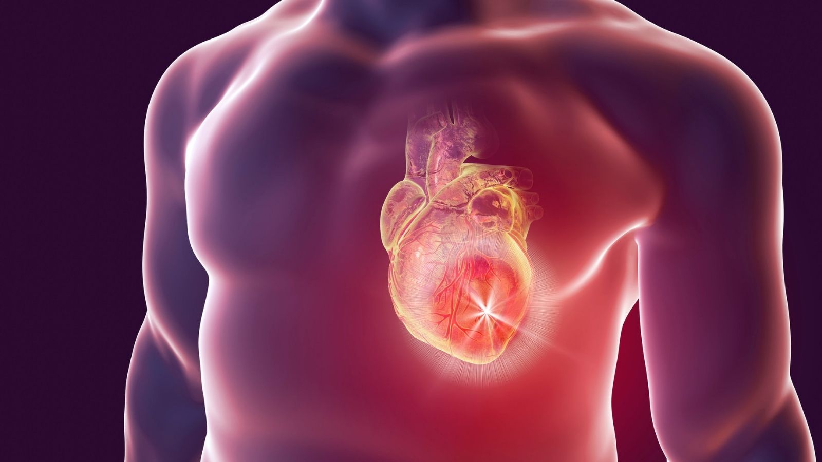
How Does the Heart’s Perfect Electrical System Work?
Our heart is actually like a two-storey house, with two chambers called “atria” (atria) on the upper floors and two more powerful and larger chambers called “ventricles” on the lower floors. The entire electrical wiring of this house is designed to ensure that blood is pumped to the right place at the right time.
This system has a main switch. Located in the upper right corner of the heart, we call this natural power source the sinoatrial (SA) node. This is your heart’s self-contained, natural pacemaker. It generates regular electrical signals about 60 to 100 times a minute, without any commands from anywhere. Like the rings created by a stone thrown into water, this signal first spreads to the chambers upstairs, the atria. This electrical impulse causes the atria to contract, emptying the blood into the ventricles downstairs. This is what the P wave, the first small and round wave we see in the ECG recording, represents.
The electrical signal then arrives at a junction between the upper and lower layers, the atrioventricular (AV) node. Here the AV node acts as a smart traffic light. It holds the signal for a very short time, measured in milliseconds. This conscious delay is vital because it allows the upper chambers to completely empty themselves of blood before the powerful chambers in the lower chambers rush to contract.
After this short pause, the signal enters a special highway system inside the heart and spreads rapidly along the walls of the ventricles. This propagation triggers a simultaneous and powerful contraction of the lower chambers. This powerful contraction is the main movement that pumps blood into the lungs and from there throughout the body. This large electrical activity is recorded as the QRS complex, the most prominent and pointed structure we see on the ECG.
Once the ventricles have completed this big job, they rest and recharge to prepare for the next heartbeat. This electrical rest and recovery phase shows up as a T wave on the ECG. In a healthy heart, this cycle continues throughout life, each beat in perfect rhythm. The ECG tells us the story of this amazing cycle.
Why is an ECG taken and which heart diseases does it give information about?
An ECG is a basic test that we use to clarify many suspicions about the heart or to rule out certain conditions. When you feel a tightness in your chest, experience palpitations such as a racing heart, feel dizzy or short of breath, an ECG is one of the first steps to find out if these symptoms have anything to do with your heart. It may also be requested as part of a general health check-up or a pre-operative assessment, even if you have no complaints.
But can an ECG detect heart disease and what conditions can this simple test tell us about? Here are some important conditions that an ECG can whisper to us:
- Heart rhythm disorders (arrhythmias)
- Heart attack (Acute Myocardial Infarction)
- An old heart attack that left a scar
- Blood supply problems in the vessels feeding the heart (Ischemia)
- Thickening or enlargement of the heart muscle (hypertrophy)
- Slowdowns in the heart’s electrical conduction system (blocks)
- Inflammation of the pericardium (pericarditis)
- Imbalances of important minerals in the body, such as potassium
- Effects of some heart medications
How to Perform a Standard ECG and What to Expect
Although the question of how to do an ECG brings to mind a complex procedure for many people, the process is actually quite simple, quick and completely painless. The entire procedure usually takes no more than 5-10 minutes, including preparation.
First, you will be asked to remove your clothes, leaving your waist and ankles uncovered. You are kindly requested to remove any metal jewelry or devices such as cell phones for a short period of time, as these may affect the signal quality.
You will then lie on your back on a comfortable examination table. It is important that you remain as still as possible and do not talk. This is because even the slightest movement of your muscles can interfere with the ECG signal and reduce the quality of the recording. You just need to keep breathing in and out calmly.
The healthcare professional performing the procedure gently wipes your skin with an alcohol swab before attaching the electrodes. This removes oil from the surface of the skin, allowing the electrodes to contact your skin and your heart to receive the electrical signals most clearly. If the hair in the chest area is dense, especially in male patients, a small area may need to be cleaned so that the electrodes can fully adhere to the skin.
A total of 10 electrodes (small, sticky scales) are then placed on your body. Four of these are glued to your arms and legs and six to specific anatomical points on your chest wall, around your heart. Each of these electrodes is like a camera looking at your heart from a different angle, allowing us to make a three-dimensional electrical map of your heart.
Once all the electrodes are in place, the technician presses the button of the ECG device. The device records the rhythm of your heart for about 10 seconds. That’s all there is to it! After the recording is finished, the electrodes are gently removed and you can immediately get dressed and go back to your daily life.
What should be considered when taking an ECG?
In order for the ECG result to be accurate and reliable, i.e. to reflect the true state of your heart, there are some simple points that both you and the staff performing the procedure should pay attention to. These rules directly affect the quality of the test.
What you need to pay attention to during the process is quite simple:
- Staying as sedentary as possible
- Avoiding talking or coughing
- Lying comfortably without straining the body
- Not crossing your legs
- Try not to shiver (indicate if you are cold)
- Not keeping the cell phone nearby
These simple precautions ensure that the ECG graph is “noise-free” and clear. Just as the model should not move when taking a photograph, your immobility is critical for us to get the clearest image. This prevents a normal ECG from being misinterpreted as problematic or a real problem from being missed.
What Do Those Lines and Waves on the ECG Mean?
When a doctor picks up an ECG printout, he or she is actually reading the chain of events inside your heart by looking at the zigzagging lines on the paper. Each peak and trough has its own meaning and represents a different stage of your heart’s electrical cycle. Although the professional answer to the question of how to read an ECG is complex, knowing what the basic waves mean can make the process more understandable.
P Wave: This is usually the first small, rounded peak at the beginning of the cycle. It shows the contraction of the atria, the upper layers of your heart. This is when the blood is mobilized to fill the ventricles, the next stop.
QRS Complex: This is the most prominent, sharpest and highest structure on the ECG. It represents the contraction of the lower chambers, the ventricles, which are the powerful pumps of your heart. This is the main event that sends blood throughout your body. The shape and width of this wave gives us information about how quickly and regularly the electricity spreads to the lower layers of the heart.
T Wave: The softer and flatter peak that follows the QRS. It shows the moment of “recovery” after a strong contraction, when the ventricles rest and re-energize for the next beat.
Your doctor will not only look for the presence of these waves. He or she will also examine their height, how long they last, their distance from each other and their shape. Together, these details provide a holistic picture of your heart’s overall health.
How to Diagnose a Heart Attack with ECG?
Yes, ECG is the fastest and most life-saving way to diagnose a heart attack in a patient who comes to the emergency room with chest pain. When one of the arteries supplying the heart (coronary arteries) is suddenly blocked by a clot, the area of the heart muscle supplied by that artery is deprived of oxygen. This state of oxygen deprivation leads to very typical changes in the electrical activity of the heart within minutes, which are immediately reflected on the ECG.
There are typical changes in the ECG during a heart attack that develop over time:
First Minutes Sometimes “hyperacute T waves” are first seen, where the T waves become much sharper and longer than normal. This is a very early finding.
Acute Phase (Minutes and Hours): The most critical finding, ST segment elevation, occurs. The ST segment, which is normally a straight line on the ECG, in this case visibly bulges upwards. This is an alarm signaling that the heart muscle has been damaged in full thickness and that the blocked vessel needs to be opened urgently by angiography. Sometimes this appearance is likened to a “tombstone” and emphasizes the seriousness of the situation.
Advanced Phase (Hours and Days): Damaged heart muscle tissue loses its electrical activity. This is manifested by the formation of deep notches on the ECG, known as pathologic Q waves. This is a sign that the damage has become permanent. At this stage, ST elevation starts to normalize.
Chronic Phase (Weeks Later): Pathologic Q waves are often permanent and remain as an “electrical scar” that a person will carry for life that they have had a heart attack in the past.
The ECG also tells us with a high degree of accuracy which wall of the heart (anterior, inferior, lateral) is affected by the attack, and therefore which main artery may be blocked. This is vital in determining our treatment strategy.
Why ECG is so Important Before and After Heart Surgery?
In a major procedure such as heart surgery, an ECG is an indispensable tool both to establish a starting point and to ensure safety throughout the journey. For this reason, ECG is routinely performed both before and after the operation.
A pre-operative ECG has two main purposes. The first is to assess the current condition of your heart and perform a risk analysis for surgery. If there are signs of a previous but unrecognized attack, an important rhythm problem or a strain in the heart muscle, we take precautions accordingly. Secondly, and perhaps most importantly, that ECG becomes your “normal”, your baseline record. When we see a change in the ECGs taken in the postoperative period, this baseline ECG gives us the answer to the question, “Is this new, or did it already exist?”. Without this possibility of comparison, it would be much more difficult to distinguish between the natural effects of surgery and a real complication.
During and after surgery, ECG monitoring is continuous in intensive care. Thanks to the monitors, we monitor every beat of your heart in real time. This uninterrupted surveillance allows us to recognize any rhythm problems that may develop due to the surgery itself or the sensitive period afterwards, or a blood supply problem in the heart muscle within seconds and intervene immediately. This follow-up is vital for the early detection and treatment of transient rhythm disturbances that we frequently encounter, especially after surgery.
What is a Stress ECG (Stress Test) and Why Is It Requested?
Sometimes, problems with the heart only become apparent when the heart has to work harder, i.e. when it is under “stress”. In particular, stenoses in the arteries supplying the heart (coronary artery disease) may not show any symptoms when the person is at rest. This is because the heart’s oxygen demand is low at rest and even a narrowed artery can somehow meet this demand. However, when you walk, jog or climb stairs, your heart’s work increases, demanding more blood and oxygen. The narrowed vessel is then unable to keep up with the increased demand and this can cause changes that can be reflected on the ECG or chest pain.
An exercise ECG (stress test) is performed to uncover this hidden problem. During the test, you walk on a treadmill or bicycle with electrodes glued to your chest at a pace that gradually increases in speed and incline. Your heart rate, blood pressure and ECG are continuously recorded.
A doctor may ask you for a stress test in cases such as
- To investigate the cause of complaints such as chest pain, tightness or shortness of breath, especially with exertion
- Assessing suspicion of asymptomatic (occult) coronary artery disease
- Safely measure a person’s exertional capacity after a heart attack or bypass surgery
- Detecting rhythm disturbances that occur only during exercise
What is Holter ECG (Rhythm Holter) and to whom is it applied?
A standard ECG is a snapshot of that 10-second moment. But what if your symptoms don’t happen at that moment? If you experience palpitations, dizziness or fainting spells that come and go only occasionally during the day, starting and ending suddenly, you are unlikely to be at the doctor at that moment. When you come to the examination, your heart may be working in a normal rhythm and the ECG taken at that time may naturally be normal.
The Holter ECG (or Rhythm Holter) is like a detective designed to catch those “elusive” moments. A Holter is a small, portable ECG recorder connected to electrodes attached to your body. You usually wear this device for 24 or 48 hours. During this time you go about all your normal daily activities, except for taking a shower; you go to work, take a walk, sleep. All the while, the device continuously records every beat of your heart.
You will be asked to write down your complaints and the time in a small notebook. When the time is up, you hand in the device and thousands of heartbeats are analyzed on the computer. Finally, your ECG recordings at the times when you report the symptoms are analyzed. This will make it clear whether your complaint is due to a rhythm problem in the heart.
Holter monitoring is particularly valuable in the following situations:
- Complaints of palpitations whose cause cannot be understood
- Unexplained episodes of fainting or feeling like fainting
- To check how effective a medication started for arrhythmia is
- To determine how often certain rhythm disturbances, such as atrial fibrillation, occur during the day.

Prof. Dr. Yavuz Beşoğul graduated from Erciyes University Faculty of Medicine in 1989 and completed his specialization in Cardiovascular Surgery in 1996. Between 1997 and 2012, he served at Eskişehir Osmangazi University Faculty of Medicine as Assistant Professor, Associate Professor, and Professor, respectively. Prof. Dr. Beşoğul, one of the pioneers of minimally invasive cardiovascular surgery in Türkiye, has specialized in closed-heart surgeries, underarm heart valve surgery, beating-heart bypass, and peripheral vascular surgery. He worked at Florence Nightingale Kızıltoprak Hospital between 2012–2014, Medicana Çamlıca Hospital between 2014–2017, and İstinye University (Medical Park) Hospital between 2017–2023. With over 100 publications and one book chapter, Prof. Dr. Beşoğul has contributed significantly to the medical literature and is known for his minimally invasive approaches that prioritize patient safety and rapid recovery.