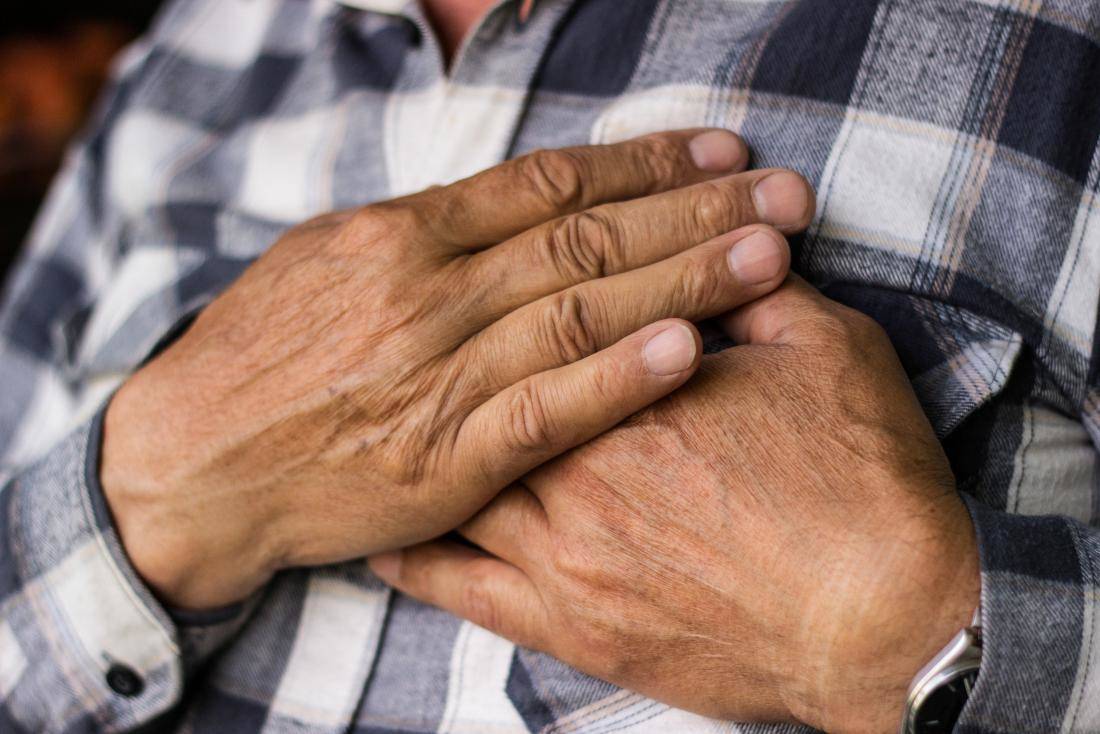
Tricuspid valve diseases involve malfunction of the valve located between the right atrium and right ventricle. Conditions such as tricuspid regurgitation or stenosis impair blood flow, leading to right-sided heart failure if untreated.
Causes include rheumatic fever, congenital malformations, endocarditis, or secondary effects of left-sided heart disease. Symptoms often manifest as fatigue, swelling in the legs, and abdominal discomfort due to fluid retention.
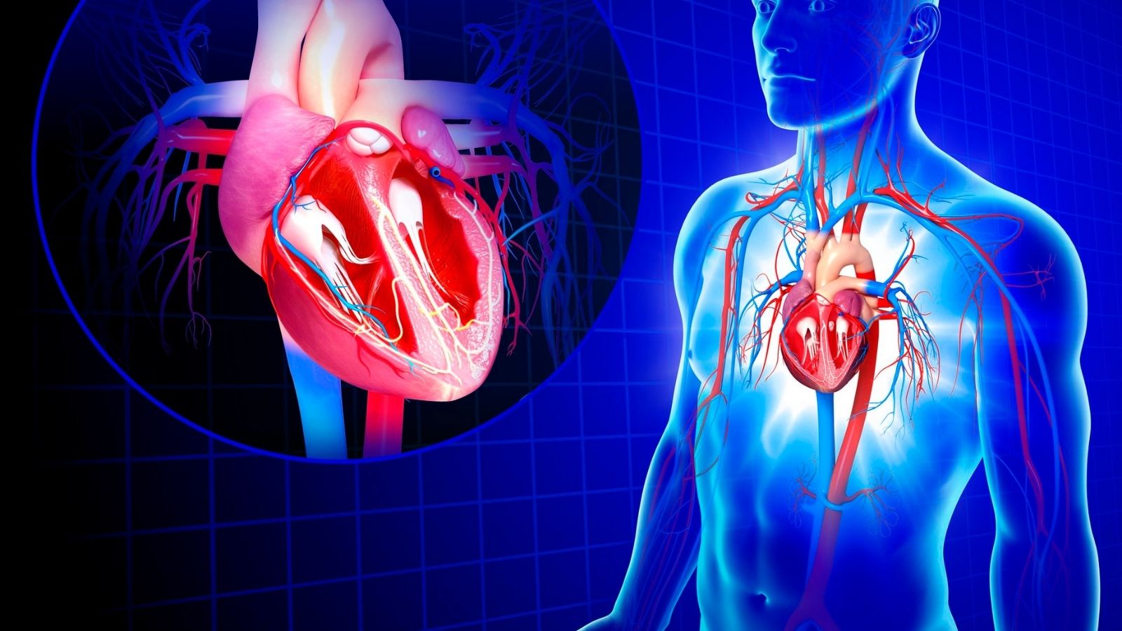
Diagnosis relies on echocardiography, which assesses valve structure and function. Additional imaging methods and cardiac catheterization may be used for detailed evaluation in complex cases.
Treatment options range from medical management with diuretics to surgical repair or valve replacement. Early intervention improves prognosis and prevents progression to advanced heart failure.
| Disease Types | – Tricuspid Stenosis
– Tricuspid Regurgitation |
| Causes | Stenosis: Rheumatic fever, congenital anomalies, carcinoid syndrome
Insufficiency: Mitral valve disease, pulmonary hypertension, endocarditis, right ventricular dilatation |
| Symptoms | Fatigue, abdominal distension (ascites), leg edema, neck vein engorgement, hepatomegaly, fatigue, jaundice, liver cirrhosis |
| Diagnostic Methods | Echocardiography (Doppler), cardiac catheterization, ECG, chest x-ray, cardiac MRI |
| Treatment Options | Diuretics, salt restriction, minimally invasive axillary and working heart surgical valve repair or replacement |
| Complications | Right heart failure, atrial fibrillation, thromboembolism, hepatic congestion, liver cirrhosis and liver failure |

Prof. Dr. Yavuz Beşoğul
>Turkey’s Cardiovascular Surgery Doctor
Why is tricuspid valve disease so important and what does it do?
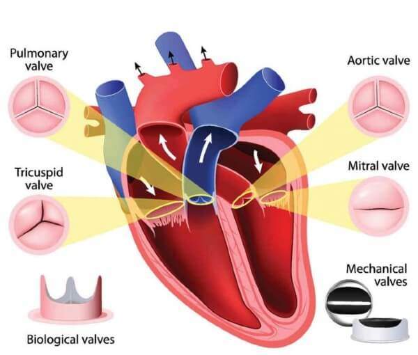
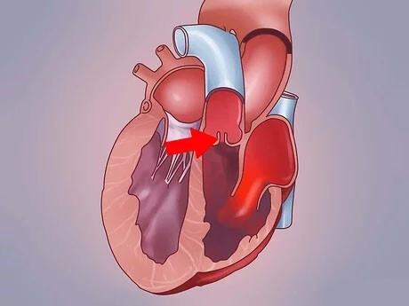
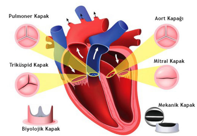
You can imagine the tricuspid valve as a highly intelligent “one-way door” between the right atrium of the heart (the first chamber where dirty blood is collected from the body) and the right ventricle (the chamber that pumps this blood to the lungs to be cleaned). It is named after the three leaflets it usually has and its main task is to manage blood flow perfectly.
When the heart relaxes, this door opens wide, allowing used and deoxygenated blood to flow freely from the right atrium into the more powerful pumping chamber below it, the right ventricle. When the right ventricle is full of blood and it is its turn to contract, this door closes tightly. In this way, the blood is pumped forward, as it should be, towards the lungs to be cleansed, and is prevented from escaping backwards, towards the atria. Thanks to this simple but vital mechanism, our heart works efficiently with every beat. When tricuspid valve disease disrupts this pattern, the load on the heart increases, the body gets tired and a number of health problems arise.
Let’s Clarify a Matter: From time to time, our patients may encounter the term “tricuspid aortic valve” and experience confusion. Having a tricuspid (i.e. three leaflets) aortic valve is a completely normal and healthy condition. The aortic valve is located on the left side of the heart and is the main valve that pumps clean blood throughout the body. In this article, we are talking about the tricuspid valve and its diseases, which is located on the right side of the heart and has a completely different role.
What types of tricuspid valve diseases are there?
The main problems we encounter with the tricuspid valve can be summarized under several main headings:
- Tricuspid Regurgitation (Back Leakage)
- Tricuspid Stenosis (Stenosis)
- Ebstein Anomaly (Congenital Problem)
- Tricuspid Atresia (Absence of the Valve)
The most common of these diseases is tricuspid regurgitation, also known as regurgitation. In this condition, the valve fails to close and when the right ventricle contracts, some of the blood leaks backwards into the right atrium instead of going to the lungs as it should. The degree of this leak is important. Very mild leaks (trace regurgitation) can occur in many healthy people and are usually not a problem. However, moderate or severe insufficiency can fatigue the heart, leading over time to enlargement of the right heart chambers and heart failure.
Tricuspid stenosis is a much rarer condition. Here the valve leaflets harden, thicken and stick together, causing the valve opening to narrow. This makes it difficult for blood to pass from the atria to the ventricles and creates a barrier.
Ebstein’s anomaly is a congenital heart defect in which the valve is incorrectly formed and positioned lower than normal. This usually leads to severe tricuspid regurgitation. Tricuspid atresia is a very rare and serious condition in which the valve never develops at birth.
What causes these tricuspid valve diseases?
The causes of these diseases are quite diverse and basically fall into two main groups: congenital and acquired.
The main congenital causes are as follows:
- Ebstein anomaly
- Tricuspid atresia
- Other complex heart defects
However, the more common acquired causes are quite varied and include some of the following:
- Left heart valve diseases
- High pulmonary blood pressure (pulmonary hypertension)
- Heart rhythm disorders (especially Atrial Fibrillation)
- Rheumatic fever
- Heart valve infection (Infective endocarditis)
- Wires from pacemaker or ICD devices
- Impacts to the chest area (Trauma)
- Rare conditions such as carcinoid syndrome
- Radiation therapy to the chest area
One point in particular should be emphasized here: The vast majority of tricuspid regurgitations are due to “secondary” causes. What does this mean? The valve itself is actually healthy, but due to a problem elsewhere in the heart (e.g. mitral valve disease on the left side or high pressure in the lungs), the right heart chambers expand over time. These enlarged chambers also stretch and tug on the ring in which the tricuspid valve sits. Because of this tension, the normally normal valves can no longer meet and close properly and leakage begins. In other words, the tricuspid valve is often an innocent victim of another disease. Therefore, treatment should not only focus on the tricuspid valve, but also address the underlying problem.
What symptoms does tricuspid valve disease cause in the body?
When the disease is in mild stages, it is often insidious and may not cause any symptoms. This is why many people can live for years without realizing they have it. But when the problem progresses and reaches moderate or serious levels, the body starts to send signals for help.
Some of the common symptoms that may occur as the disease progresses include
- Persistent fatigue and weakness
- Shortness of breath, especially with movement
- Swelling in the feet, legs and abdomen (edema)
- Palpitations or irregular heartbeat
- Visible pulsation or twitching sensation in the neck veins
- Feeling of fullness and tenderness in the right upper abdomen
- Loss of appetite
- Coldness of the skin, especially hands and feet
Simply put, shortness of breath and fatigue are caused by the heart’s inability to pump enough blood efficiently around the body. Swelling in the legs or abdomen (edema) occurs when the right heart is unable to push the blood forward and the blood pools in the veins and fluid leaks out of the veins. The marked pulsation in the veins of the neck is a reflection of the pressure wave created by the backward blood flow. If you experience one or more of these symptoms, it is best to consult a specialist without neglecting the situation.
How is tricuspid valve disease diagnosed?
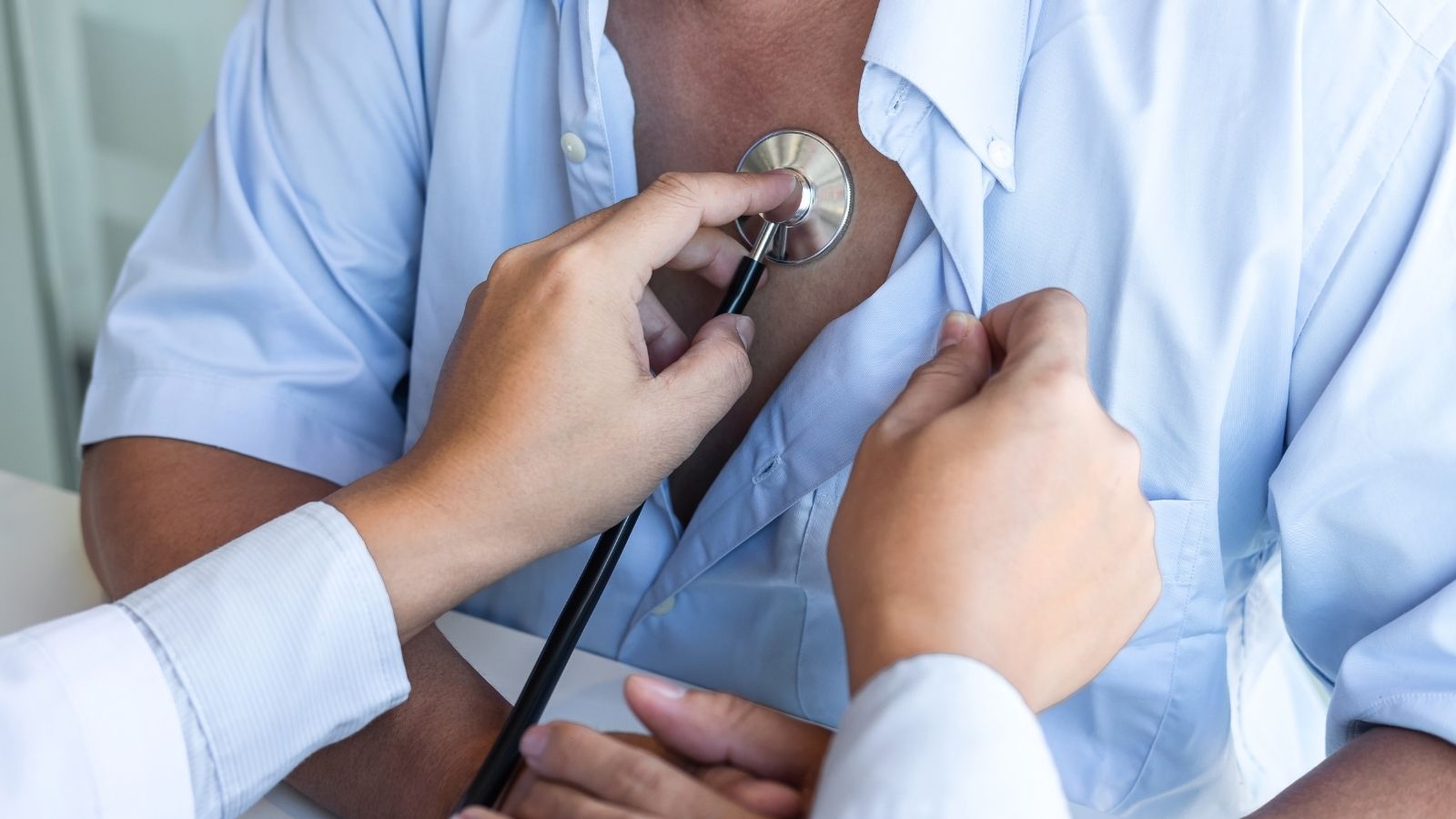
Making a correct diagnosis is the most important first step in our treatment journey. This process starts with a detailed interview with you. We listen to your complaints, when they started and how they affect your life. In the physical examination, we will then listen to your heart with a stethoscope to check for abnormal sounds called murmurs, fullness in your neck veins or edema in your legs.
After this initial assessment, there are a number of basic and advanced tests that we use to confirm the diagnosis and clarify the severity of the disease:
- Echocardiography (ECHO – Heart Ultrasound)
- Electrocardiogram (ECG)
- Chest X-ray
- Cardiac Magnetic Resonance (Cardiac MRI)
- Exercise Stress Test (Effort Test)
- Cardiac Catheterization (Angiography)
Among these tests, Echocardiography (ECHO) is the gold standard for diagnosing tricuspid valve disease. ECHO is a painless and harmless ultrasound procedure that provides live, moving images of your heart and valves. In these images, we can clearly see the structure of the valve, how the leaflets move, whether there is a leak or stenosis in the valve, and if so, to what extent. We also measure how much the chambers on the right side of the heart are enlarged and the strength of contraction. Sometimes we can resort to Transesophageal Echocardiography (TEE) through the esophagus to get even clearer images.
Other tests help us to complete the picture. We can evaluate the heart rhythm with ECG, the overall size of the heart with chest X-ray, and the functions of the right ventricle in particular in much more detail with Cardiac MRI.
What treatment options are currently available for tricuspid valve disease?
The treatment of your tricuspid valve disease is tailored to you, like a tailor making a dress just for you. Many factors, such as the type of disease, its severity, your age, general health and lifestyle, will determine which treatment pathway we choose. Our main goal in treatment is to relieve your symptoms, prevent further strain on your heart and give you a good quality of life.
Medication and Lifestyle Modifications:
This approach is usually the first step in treatment. Medicines do not eliminate the disease, but control symptoms by making the heart’s work easier. Diuretics (diuretics) reduce edema by removing excess fluid from the body. Rhythm regulators control palpitations. Blood thinners prevent clot formation, especially if accompanied by arrhythmia. In addition, reducing salt consumption, eating a healthy diet and being as active as your doctor allows are essential for treatment. However, continuing medication for a long time can lead to cardiac cirrhosis and lose the chance of surgery. Surgical treatment should be recommended before liver destruction begins.
Surgical Treatment Repairing or Renewing the Door of the Heart
When medication is inadequate or the valve problem is severe, we turn to more permanent solutions. The timing of surgery is very important. An intervention at the right time, before irreversible damage to the heart occurs, can be life-saving.
When surgery is needed, there are two main approaches:
- Tricuspid Valve Repair
- Tricuspid Valve Replacement
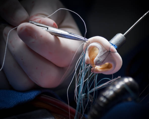
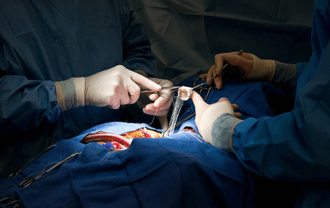
Whenever possible, our first choice is to repair the patient’s own valve. Because preserving your own tissue is always the healthiest. The most common repair technique we use is to narrow the enlarged valve ring to the required size with a special ring (annuloplasty). This allows the valves to close properly again.
But sometimes the valve is so damaged that it cannot be repaired. In this case, the damaged valve is removed and replaced with either a biological valve (made of animal heart membrane) or a mechanical valve (made of metal and carbon). Nowadays, these operations are minimally invasive, performed through a small incision in the armpit while the heart is working. This has the advantage of preventing heart rhythm block.
Catheter Methods Non-Surgical Modern Solutions
One of the most exciting developments in the world of medicine in recent years has been the development of catheter-based techniques for patients who are at high risk of or unable to undergo heart surgery. In these techniques, a thin tube (catheter) is usually inserted into the heart through a vein in the groin and the entire procedure is performed from there. Today, however, the chances of success are still not high.
What is the recovery period after treatment of tricuspid valve diseases?
Your recovery will depend on the type of treatment you have received. While recovery after open heart surgery can take several months, it is much shorter after minimally invasive surgery or catheterization.
Regardless of the method chosen, cardiac rehabilitation programs are the most important supporter of recovery. These programs help you return to life with confidence by strengthening not only your body but also your spirit with exercises, nutrition education and psychological support tailored to you.
Here are some general points to consider during the recovery period:
- Using the medicines prescribed by your doctor without interruption
- Careful wound care if you have had surgery
- Avoid heavy lifting for the specified period
- Not driving until your doctor gives permission
- Participating in your recommended cardiac rehabilitation program
- Not to disrupt the follow-up and control appointments
What should I consider when living with tricuspid valve disease?
With this diagnosis, some positive changes in your life will both protect your heart and improve your overall quality of life. Your biggest helper in this process will be yourself.
Paying attention to the following points in your diet will ease the burden on your heart:
- Reduce salt (especially if there is edema)
- Avoiding packaged and processed foods
- Including plenty of vegetables and fruits on your table every day
- Prefer healthy fats (olive oil, avocado, walnuts)
- Drink enough water (unless your doctor has recommended fluid restriction)
- Limit sugary and pastry foods
In particular, protection against heart valve infection (infective endocarditis) is vital:
- Taking great care of your oral and dental health
- Visiting the dentist at least twice a year
- Always inform your cardiologist before dental treatments or other surgical procedures with a risk of bleeding
- Getting any infection that develops in your body (urinary tract, skin, etc.) treated immediately
- Flu and pneumonia vaccinations as recommended by your doctor
Tricuspid valve disease is no longer a helpless or “forgotten” problem. Thanks to early diagnosis, correct timing and early surgery before the onset of cardiac cirrhosis, it is possible to overcome these diseases and live a healthy, quality life.
After Closed Tricuspid Valve Surgery
| Type of Surgery | Minimally invasive tricuspid valve repair (annuloplasty) or valve replacement (mechanical or biological). |
| Recovery Time | Average 10-20 days, much faster than classical open surgery since no sternotomy is performed. |
| Medication Use | Warfarin (in mechanical valve), rhythm regulators, diuretics and heart failure medications according to valve type. |
| INR Monitoring | Lifelong in case of a mechanical valve, not required in case of a biological valve. |
| Physical Activity | Early mobilization with light walking is recommended; heavy exercise should be avoided for 15 days. |
| Rhythm and Pulse Monitoring | Rhythm disorders such as AV block and atrial fibrillation may be observed after tricuspid valve surgery. |
| Imaging and Control | Echocardiography should monitor valve function, right ventricular performance and pulmonary pressures. |
| Complications | Risk of valvular dysfunction, arrhythmia, pericarditis, stroke, pleural effusion, right heart failure. |
| Wound Care | after 10 days you don’t need |
| Nutrition | Salt restriction (to reduce edema), low-fat, heart-friendly diet is recommended. |
| Smoking and Alcohol | Not recommended, especially in the presence of pulmonary hypertension or right heart failure. |
| Sexual Activity | It can usually be started within 2-4 weeks once general well-being is achieved. |
| Psychological Support | Psychological support is useful in patients with chronic right-sided heart failure and rhythm problems. |
| Vehicle Use | It is usually possible after 10-15 days when reflexes and concentration are in order. |
| Controls | Echocardiography is recommended once a month for the first 3 months, then every 3-6 months and annually thereafter. |

Prof. Dr. Yavuz Beşoğul graduated from Erciyes University Faculty of Medicine in 1989 and completed his specialization in Cardiovascular Surgery in 1996. Between 1997 and 2012, he served at Eskişehir Osmangazi University Faculty of Medicine as Assistant Professor, Associate Professor, and Professor, respectively. Prof. Dr. Beşoğul, one of the pioneers of minimally invasive cardiovascular surgery in Türkiye, has specialized in closed-heart surgeries, underarm heart valve surgery, beating-heart bypass, and peripheral vascular surgery. He worked at Florence Nightingale Kızıltoprak Hospital between 2012–2014, Medicana Çamlıca Hospital between 2014–2017, and İstinye University (Medical Park) Hospital between 2017–2023. With over 100 publications and one book chapter, Prof. Dr. Beşoğul has contributed significantly to the medical literature and is known for his minimally invasive approaches that prioritize patient safety and rapid recovery.