A leaky heart valve, medically termed valvular regurgitation, occurs when a valve fails to close properly, allowing blood to flow backward. This condition can strain the heart, leading to symptoms such as fatigue, shortness of breath, and edema.
Types of valvular regurgitation include mitral, aortic, tricuspid, and pulmonary insufficiency. Each type has distinct causes, such as rheumatic disease, congenital defects, or age-related degeneration, requiring tailored evaluation.
Diagnostic tools like echocardiography and cardiac MRI assess valve structure, regurgitation severity, and impact on heart function. Early detection is crucial to prevent heart failure and associated complications.
Treatment may involve medical management with diuretics and vasodilators, or surgical repair and replacement in advanced cases. Regular monitoring ensures optimal timing of intervention and long-term cardiac stability.
| Mitral Valve Leakage | Causes: Mitral prolapse, endocarditis, rheumatic disease, left ventricular dilatation
Symptoms: Shortness of breath, palpitations, orthopnea, leg edema Diagnosis: ECHO (transthoracic/transesophageal), ECG, chest radiography Treatment: Medical (ACEi, diuretics), surgical valve repair or replacement Complications: Left ventricular dilatation, AF, heart failure |
| Aortic Valve Leakage | Causes: Degenerative change, bicuspid valve, endocarditis, rheumatic disease
Symptoms: Exercise intolerance, chest pain, fainting, dyspnea Diagnosis: ECHO, CT/MR, aortic angiography Treatment: Medical monitoring, surgical valve replacement, TAVI Complications: Left ventricular hypertrophy, heart failure, sudden death |
| Tricuspid Valve Leakage | Causes: Right ventricular failure, pulmonary hypertension, endocarditis
Symptoms: Neck venous engorgement, leg edema, liver enlargement Diagnosis: ECHO, liver tests Treatment: Diuretics, surgical or percutaneous repair Complications: Right heart failure, hepatomegaly |
| Pulmonary Valve Leakage | Causes: Congenital defect, carcinoid syndrome
Symptoms: Shortness of breath, exercise intolerance, palpitations Diagnosis ECHO, MRI Treatment Medical follow-up, rarely surgical intervention Complications: Right ventricular dilatation, right heart failure |
What is a Heart Valve Leak?
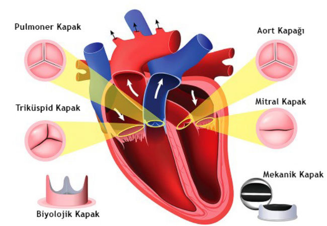
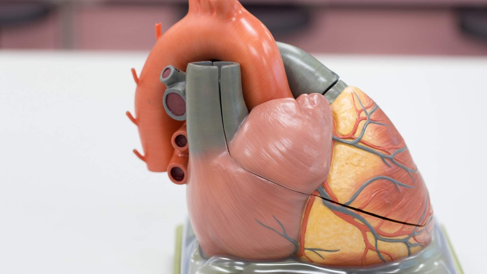
Inside our heart, there are four valves that perfectly manage the flow of blood. You can think of them as doors in a building that only open in one direction. Their job is to make sure that blood never backs up as it passes between the chambers and from the heart to the main arteries. On the right side of the heart are the tricuspid and pulmonary valves, and on the left side are the mitral and aortic valves. With every heartbeat, these valves open and close in perfect timing.
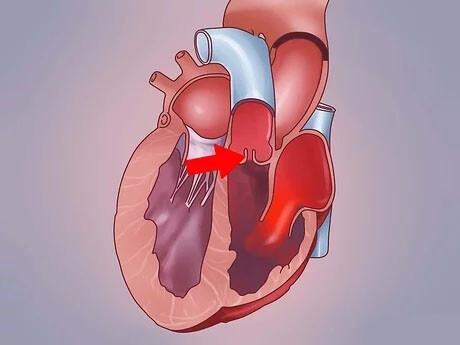
When the heart valve leaks, this one-way door system breaks down. The valves cannot close completely and tightly, leaving a gap around the edges. When the heart contracts and tries to pump blood forward, some blood leaks backwards through this gap into the chamber from which it came. In medical language, this is called “regurgitation” or “regurgitation”. It is called a deficiency because the valve is not able to do its job of holding back the blood.
The most important consequence of this backflow is that it creates “volume overload” in the heart. With each beat, the heart chamber from which the blood has backed up has to accommodate both the blood coming in normally and the blood leaking through the valve. For example, in an aortic valve leak, after the blood is ejected into the aorta for distribution throughout the body, some of it returns to the left ventricle, the main pumping chamber of the heart. This causes the left ventricle to expand (like a balloon inflating) and thicken its wall over time to cope with the extra blood. To compensate, the heart tries to contract more strongly. This adaptation process is called “cardiac remodeling” or remodeling of the heart.
Initially, this adaptive effort of the heart prevents the person from feeling any problems. However, over the years, this constant overtime fatigues the heart. Just as a rubber band that is constantly stretched and left unstretched loses its elasticity and strength over time, the heart muscle loses its elasticity, weakens and eventually becomes unable to effectively pump the blood the body needs. This final stage is known as heart failure and is one of the most serious consequences of a valve leak. The popular expression “the heart valve is leaking air” is a complete misconception; it is not the air that leaks, but the blood itself.
What are the Symptoms of Heart Valve Leakage?
Leakage of the heart valve can be quite insidious, especially when it is mild, and can be asymptomatic for years. For this reason, many people learn about a mild heart valve leak by chance during a check-up for another reason. However, when the leakage reaches moderate or severe levels, the increased workload of the heart begins to reflect on the body and various symptoms appear. These heart valve leakage symptoms can vary depending on which valve the leak is in and how severe it is.
The most common symptoms are:
- Shortness of breath
- Fatigue and quick fatigue
- Palpitations
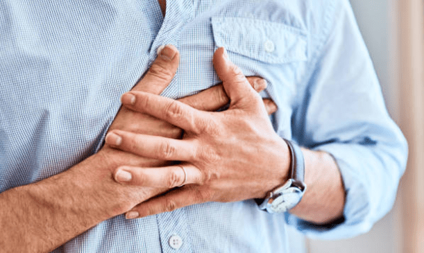
- Discomfort in the chest
- Swelling of the feet and legs
- Dizziness or fainting
- Persistent cough
- Abdominal bloating
Let us examine what these symptoms mean in a little more detail. Shortness of breath is perhaps the most important and most common symptom. It occurs especially when climbing stairs, climbing hills or walking briskly. As the condition progresses, you may feel as if you are short of breath while walking in a straight line or even while resting. Waking up at night with shortness of breath or needing to lie down with several pillows to breathe easily may indicate that the problem is getting serious. The main cause of this condition is the accumulation of blood in the lungs from valve leaks on the left side of the heart (mitral or aortic).
Fatigue and fatigue are also very common. Because there is not enough oxygen-rich blood supply to the body tissues, the person feels constantly drained of energy and exhausted. The things he used to do easily start to grow on him.
Palpitations are when you feel your heart beating differently from normal. This can range from a mild sensation, such as the “flapping of a bird’s wing”, to a strong and irregular beating of the heart, as if it is about to burst out of the chest. This is usually a sign of rhythm disturbances, such as atrial fibrillation, which develops due to a valve problem and should be taken seriously.
Swelling of the feet, ankles and legs (edema) is more common with leaky valves, especially on the right side of the heart (tricuspid). When the heart is unable to collect and pump blood efficiently, the blood pools in the veins in the legs and fluid begins to leak between the tissues. This swelling, which initially appears at the end of the day, may become permanent over time.
Dizziness and fainting is an alarming symptom, especially in severe aortic valve leaks. It is caused by insufficient blood flow to the brain and can occur with sudden changes in position or during exertion. This requires urgent medical evaluation.
What Causes Heart Valve Leakage?
So what are the factors that prevent these doors from closing properly, i.e. what triggers the question of why does the heart leak? There is no single answer to the question of why a heart valve leaks; there can be many different underlying causes. In some cases, the structure of the valve is congenitally defective, while in other cases it is damaged over time or as a result of another disease.
The main reasons are as follows:
- Mitral valve prolapse (MVP)
- Calcification and wear and tear due to aging (degeneration)
- Rheumatic heart disease
- Previous heart attack
- Congenital heart defects
- Diseases of the heart muscle (cardiomyopathy)
- Valve inflammation (infective endocarditis)
- Uncontrolled high blood pressure (hypertension)
- Connective tissue diseases such as Marfan syndrome
- Radiotherapy to the chest area
- Serious chest trauma
mitral valve prolapse is a condition in which the leaflets of the mitral valve are looser and more flexible than normal. When the heart contracts, these leaflets bulge backwards into the left atrium like a sailboat, preventing full closure and causing leakage. This is a fairly common condition in the community and is usually benign, but in some people it can progress to serious leakage over time.
Aging inevitably leads to a wear and tear of our tissues. Heart valves are also affected by this process. Over the years, calcium can build up in the valves (calcification), their tissues can harden and lose elasticity. These degenerative changes are the most common cause of aortic and mitral valve leaks, especially in people over the age of 65.
Rheumatic heart disease used to be the most important cause of valvular disease, but has declined with the widespread use of antibiotics. However, it is still a serious risk in developing countries and in people who had untreated beta germ throat infections in childhood. This disease causes inflammation of the valves and permanent damage that occurs years later.
A heart attack can also impair valve function. During a heart attack, part of the heart muscle dies. If this dead muscle tissue contains the “papillary muscles” that hold the valves in place and work like the strings of a parachute, these structures lose their function and the valve can suddenly and violently start to leak. This is a life-threatening situation that requires urgent intervention.
Finally, some people are born with congenital valve anomalies. For example, while the aortic valve normally has three leaflets, it is common for some people to have two leaflets (bicuspid aorta). As this two-leaflet valve is subjected to more stress than normal, it wears out, calcifies and can start to leak at an earlier age.
How Is Heart Valve Leakage Detected?
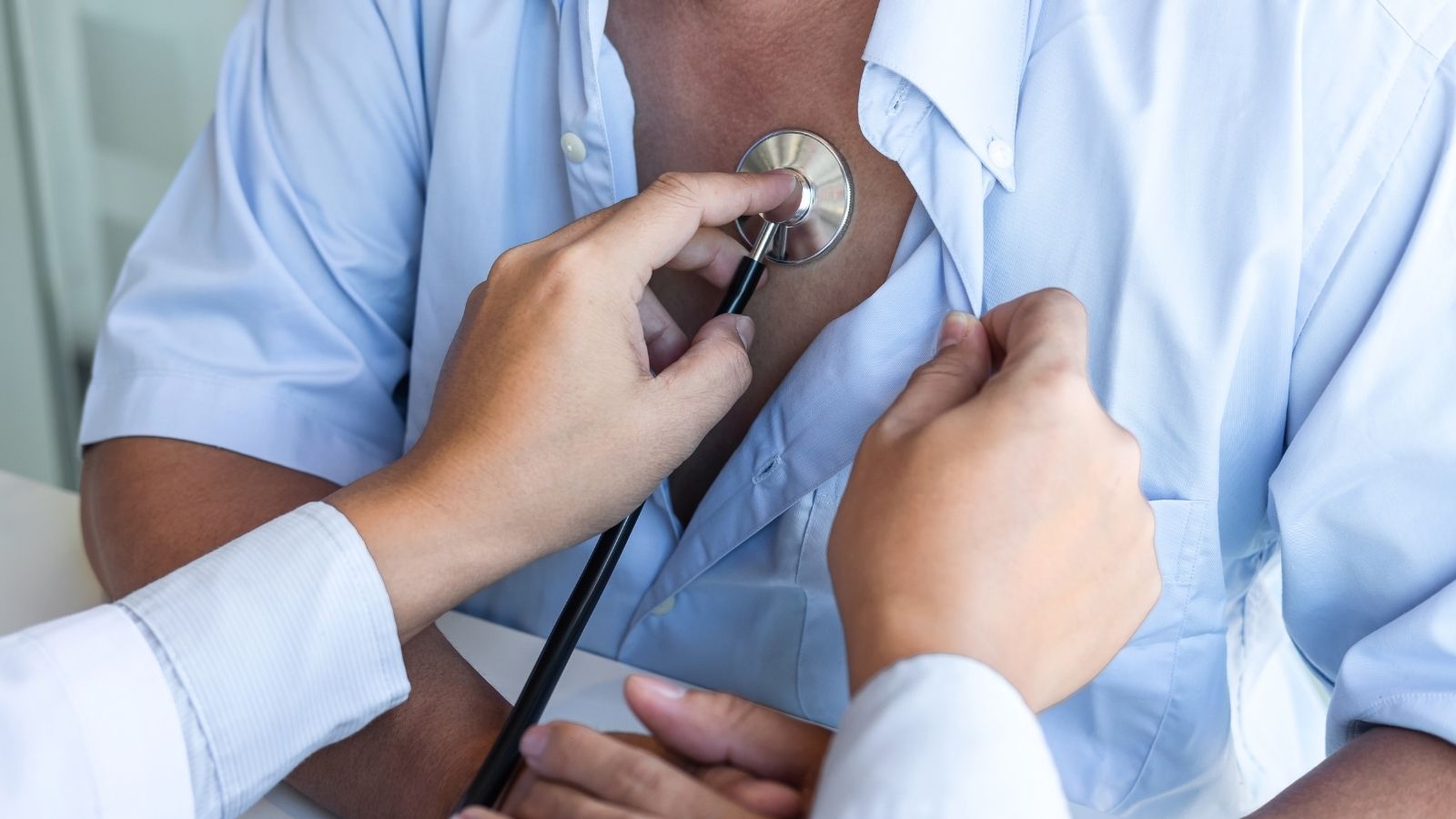
When you consult a physician with a suspected leaky heart valve, the diagnostic process involves several steps. It starts with listening to your story. Details such as your complaints, when they started and what triggered them are very important. A thorough physical examination can then provide one of the most critical diagnostic clues. When the physician listens to your heart with a stethoscope, he or she may hear a “murmur”. A murmur is an abnormal, whispering or buzzing sound created by blood as it passes through a leaky valve. When this sound is heard, its intensity and in which area of the heart it is clearest can give important information about which valve may have a problem.
However, a murmur is only a suspicion; the most valuable test to confirm the diagnosis and chart the course of the disease is Echocardiography (ECHO), an ultrasound of the heart. This test uses sound waves to create instant, moving images of your heart. It is painless, does not involve radiation and is the gold standard method for evaluating a heart valve disease.
Echocardiography provides the following information:
- Which valve or valves are leaking
- Severity of leakage (mild, moderate, severe)
- Structure of the valves (thickening, calcification, whether there is a tear)
- The size of the heart chambers (whether dilated or not)
- Thickness of the heart walls
- The pumping power of the heart (Ejection Fraction – EF)
Usually, Transthoracic Echocardiography (TTE) performed through the chest wall is sufficient. However, in some cases, especially when a clear image cannot be obtained due to the patient’s chest structure or lung problems, or when the valve structure is to be examined in more detail, a method called Transesophageal Echocardiography (TEE) is used. In this method, a thin tube with a small ultrasound camera at the end is lowered into the esophagus by having the patient swallow it under mild anesthesia. Since the esophagus is located just behind the heart, this method provides much clearer and more detailed images.
In addition to these basic tests, other tests may be ordered on a case-by-case basis to provide additional information. An electrocardiogram (ECG) records the electrical activity of the heart and detects possible rhythm disturbances. A chest x-ray gives an idea of the overall size of the heart and whether there is fluid accumulation in the lungs. An effort test (stress test) is used in patients with vague symptoms to see if complaints occur during exertion and the heart’s response to the load. Cardiac Magnetic Resonance (Cardiac MRI) is an advanced imaging modality used to measure the amount of leakage very precisely and to assess the condition of the heart muscle in detail.
Is Heart Valve Leakage Life Threatening If Left Untreated?
This is one of the questions that patients and their relatives are most concerned about and ask most often: “Is heart valve disease fatal?” The answer to this question depends on the severity of the condition and the timely intervention. A mild leak in the heart valve is usually not life-threatening and does not affect a person’s life expectancy. These patients are usually only followed up regularly.
However, if left untreated, moderate and especially severe heart valve leakage can lead to a series of serious and potentially life-threatening problems by disrupting the structure and function of the heart. If the heart is constantly working at a higher than normal load, over time the compensatory mechanisms become inadequate.
The main dangerous consequences of a serious untreated valve leak are the following:
- Heart failure
- Atrial fibrillation and related stroke
- Pulmonary hypertension (elevated lung pressure)
- Infective endocarditis (valve inflammation)
- Sudden cardiac death
The most common and most important of these complications is heart failure. The heart muscle, which has been overworked for years, eventually tires and is unable to perform its function as a pump effectively. This condition manifests itself with symptoms such as shortness of breath, fluid accumulation in the body (edema) and extreme fatigue and severely reduces the patient’s quality of life.
Another important risk is a rhythm disorder called atrial fibrillation. When the left atrium of the heart enlarges, especially due to mitral valve leakage, the normal electrical pattern of this chamber is disrupted and it begins to beat irregularly and rapidly. The most dangerous consequence of atrial fibrillation is that blood pools in the heart and forms a clot. If this clot breaks off and blocks the brain vessels, it can result in a stroke.
Left-sided valve leaks (mitral and aortic) cause blood to constantly backpressure into the lungs, raising the pressure in the pulmonary arteries. This is called pulmonary hypertension and can worsen shortness of breath and lead to failure of the right side of the heart.
Damaged valve surfaces create an environment where bacteria entering the bloodstream can easily settle and multiply. This increases the risk of infective endocarditis, or valve inflammation. This is a serious infection that causes further damage to the valve and requires urgent treatment.
Although rare, especially in cases of advanced and symptomatic aortic valve insufficiency, fatal rhythm disturbances can be triggered and the risk of sudden cardiac death can occur.
What Treatment Methods Are Used for Heart Valve Leakage?
Heart valve leak treatment is not a “one-size-fits-all” prescription. The treatment plan is completely individualized depending on which valve the leak is in, its severity, the presence of symptoms, the extent to which the heart is affected, and the patient’s age and general health. The main goal is to relieve symptoms, improve quality of life and, most importantly, prolong life by solving the problem before irreversible damage to the heart occurs.
Observation and Medication
Not all heart valve leaks require immediate surgery. For mild or asymptomatic moderate leaks, an approach called “watchful waiting” or “medical follow-up” is usually adopted. During this period, the patient is checked at regular intervals (usually every 6 months or year) with echocardiography and a medical examination.
Medication does not mechanically correct the leak in the valve, i.e. it does not eliminate the leak. However, it is extremely important to temporarily control symptoms and ease the workload of the heart. The main groups of drugs used for this purpose are:
- Drugs that lower blood pressure and make it easier for the heart to pump blood (ACE inhibitors, ARBs)
- Drugs that slow the heart rate and make the heart work more efficiently and regulate blood pressure (Beta blockers)
- Drugs that reduce edema and shortness of breath by removing excess fluid from the body (diuretics or diuretics)
- Drugs to control palpitations or arrhythmia if it has developed (antiarrhythmics)
- Blood thinners (anticoagulants) used to reduce the risk of clots are vital, especially in patients who develop atrial fibrillation.
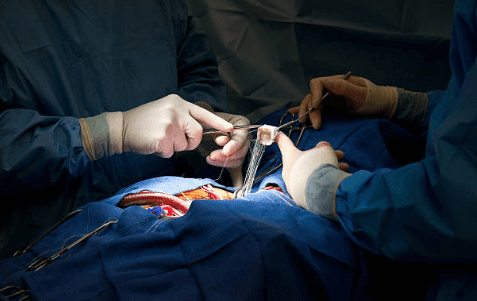
Surgical Treatment
Surgical intervention is the permanent solution when medication is inadequate, when the leak has reached an advanced stage, when symptoms reduce the patient’s quality of life, or when echocardiography shows deterioration in the function of the heart even in the absence of symptoms. There are two main surgical options for treating heart leakage: cover repair or cover replacement.
Heart Valve Repair: It is the first and most preferred method for surgeons whenever possible. In this method, the patient’s own valve tissue is preserved and the problem causing the leak is corrected. For example, an enlarged valve ring can be narrowed with a special ring, a sagging or torn valve leaflet can be corrected, or broken valve strands (chordae) can be repaired. The biggest advantages of valve repair are that the body continues to live with its own tissue, there is usually no need to use blood thinners for life, the risk of infection is lower and life expectancy is better in the long term.
Heart Valve Replacement: If the valve tissue is damaged beyond repair due to infection, excessive calcification or rheumatic disease, the damaged valve is removed and replaced with an artificial (prosthetic) valve.
There are two main types of prosthetic valve:
- Mechanical covers
- Biological (tissue) closures
Mechanical closures are made of highly durable materials such as titanium or carbon and theoretically last a lifetime. However, the biggest disadvantage is the risk of clots forming on their surface. Therefore, patients with mechanical valve implantation must take effective blood thinners such as Coumadin (warfarin) regularly for the rest of their lives to eliminate this risk and must be monitored continuously with blood tests (INR).
Biological valves are usually made from specially treated heart tissue (pericardium) from cattle or pigs. Their most important advantage is that they have a very low risk of clot formation and therefore usually do not require long-term use of blood thinners. However, these valves are not as durable as mechanical valves and wear out over time. Their average lifespan is around 15-20 years and at the end of this period, they may need to be replaced with another surgery. For this reason, they are generally preferred in older patients or those who cannot use blood thinners.
Modern Minimally Invasive and Catheter Methods
Today, the treatment of heart valve leakage is not limited to classical open heart surgery in which the sternum is completely cut. With techniques called minimally invasive surgery or closed heart surgery, valve repair or replacement can be successfully performed through much smaller incisions made in the side wall of the chest (armpit) or under the breast. These methods offer less bleeding, less pain, less bleeding, faster recovery and better cosmetic results. In elderly patients or patients with additional serious health problems who are too high risk for open heart surgery, non-surgical catheterization through an artery in the groin offers hope. For example, in suitable patients with severe mitral valve leakage, a latch system called “MitraClip” is delivered to the heart via catheter and the leaky leaflets of the valve are pinned together to reduce leakage. These methods play an important role in improving the quality of life of patients by eliminating the risks of surgery.
Frequently Asked Questions
What is a heart valve leak?
It is a condition in which the heart valves do not close properly and blood leaks backwards. The medical name is “valve insufficiency”.
Which heart valves can leak?
Leakage may develop in mitral, aortic, tricuspid and pulmonary valves. The mitral and aortic valves are most commonly affected.
What are the symptoms of a heart valve leak?
Symptoms may include shortness of breath, fatigue, palpitations, chest pain, swelling in the legs and fainting.
Is leakage always symptomatic?
No. Mild leaks are usually asymptomatic and can be detected during routine check-ups.
Why does a heart valve leak occur?
Congenital causes, rheumatic diseases, aging, infection (endocarditis), heart enlargement and trauma are the main causes.
Will the fugitive advance?
Warning. Over time, a mild leak can become moderate or severe and impair the function of the heart.
How is it diagnosed?
The most effective diagnostic method is echocardiography (ECHO). The backflow of blood through the valve is clearly seen with this method.
Does leakage cause enlarged heart?
Yes, this is true. As the leakage increases, the heart has to work harder and may develop enlargement (dilatation) over time.
Does every leak require treatment?
No, no, no. Mild cases are only monitored. Moderate and severe cases are treated.
Is medication possible?
Medicines are used to relieve symptoms but do not completely eliminate the leak. In some cases surgery may be required.
Do I need surgery?
If there is severe leakage and heart function is affected, valve repair or valve replacement surgery is recommended.
Is cover repair or cover replacement better?
If possible, valve repair is preferred. However, very damaged valves are replaced with mechanical or biological valves.
Is it possible to prevent leakage progression?
Progression can be slowed with blood pressure control, protection against infections, regular follow-up and medication.
Can a person with a leak exercise?
People with mild leakage can do controlled exercise. Moderate and severe leaks require treatment.

Prof. Dr. Yavuz Beşoğul graduated from Erciyes University Faculty of Medicine in 1989 and completed his specialization in Cardiovascular Surgery in 1996. Between 1997 and 2012, he served at Eskişehir Osmangazi University Faculty of Medicine as Assistant Professor, Associate Professor, and Professor, respectively. Prof. Dr. Beşoğul, one of the pioneers of minimally invasive cardiovascular surgery in Türkiye, has specialized in closed-heart surgeries, underarm heart valve surgery, beating-heart bypass, and peripheral vascular surgery. He worked at Florence Nightingale Kızıltoprak Hospital between 2012–2014, Medicana Çamlıca Hospital between 2014–2017, and İstinye University (Medical Park) Hospital between 2017–2023. With over 100 publications and one book chapter, Prof. Dr. Beşoğul has contributed significantly to the medical literature and is known for his minimally invasive approaches that prioritize patient safety and rapid recovery.