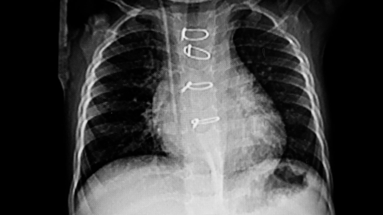
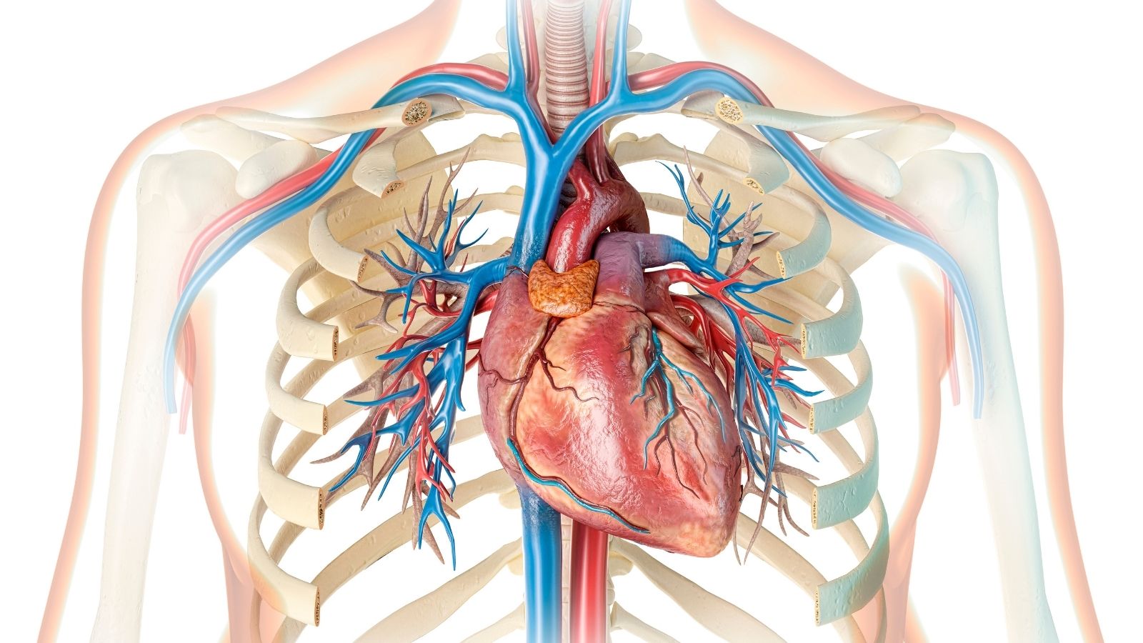
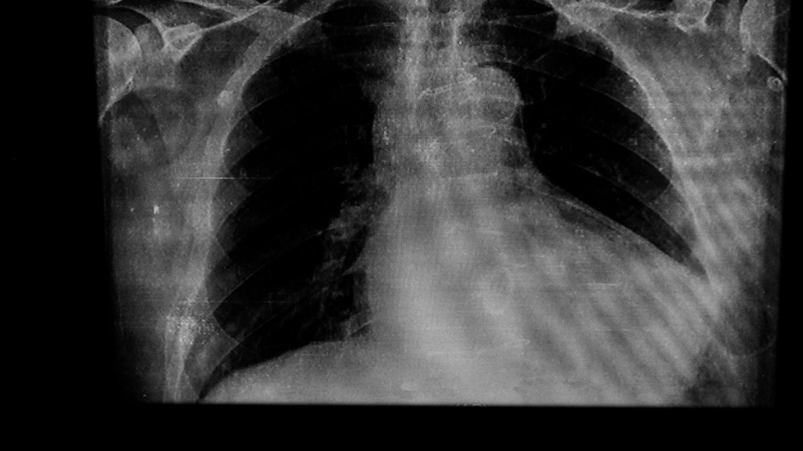
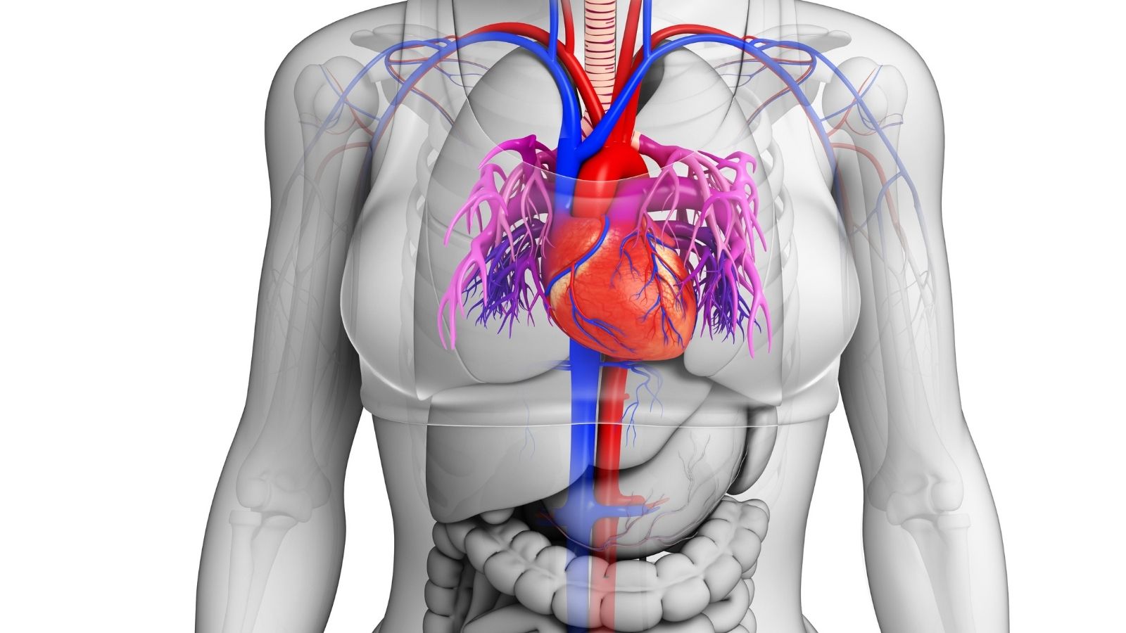
CT coronary angiography is a non-invasive imaging method used to visualize coronary arteries. It provides detailed information about blockages and helps in diagnosing coronary artery disease.
The procedure involves intravenous contrast injection and high-resolution CT scanning, allowing precise assessment of arterial narrowing without the need for catheterization.
CT angiography is highly valuable in detecting early-stage atherosclerosis, evaluating chest pain, and planning treatment. It reduces the need for invasive diagnostic procedures.
Safety measures, such as minimizing radiation exposure and checking kidney function before contrast use, are applied to ensure patient well-being during CT coronary angiography.
| Medical Name | Computed Tomography Coronary Angiography (CT Coronary Angiography, Virtual Angiography) |
| Common Uses | – Evaluation of vessels in patients with suspected coronary artery disease – Detection of vessel occlusion and stenosis |
| Causes | – Chest pain and risk of cardiovascular disease – Request for non-invasive evaluation as an alternative to conventional angiography |
| Risk Factors | – Allergy to contrast media – Impaired renal function – Radiation sensitivity (pregnancy, etc.) |
| Complications | – Allergic reaction due to contrast media- Temporary or permanent impairment of renal function- Radiation exposure |
| Diagnostic Methods | – Preliminary clinical evaluation – Blood tests (especially creatinine for renal function, etc.) – ECG, other cardiologic tests if necessary |
| Treatment Methods | – CT coronary angiography is used for diagnostic purposes and is not a treatment method |
| Prevention Methods | – Control of renal function before contrast media- Taking precautions in those with a history of allergy- Avoiding unnecessary radiation exposure |
What Are the Main Differences Between Virtual Angiography and Classic Angiography?
One of the most common questions patients ask is how these two methods differ from each other. The main distinction between them is the way the procedure is performed and the patient’s experience of the process.
CT Coronary Angiography (Virtual Angiography) is a completely external imaging test. During the procedure, a special dye called “contrast medium” is injected into a vein in your arm, one of the veins through which we give blood. This material enters your bloodstream and reaches your cardiac arteries. At this point, while you lie on the bed of the CT scanner, it takes hundreds of slices of your heart in a few seconds to complete the image. No catheters, wires or other medical instruments are inserted into your body, especially into your arteries, during the process. This makes virtual angiography a non-invasive, painless and much more comfortable option for the patient.
Classical (Invasive) Coronary Angiography, as the name suggests, is an invasive procedure, meaning that it is performed by entering the body. It is usually performed in a hospital setting, in a special room called a “catheter laboratory”. An artery in the groin or wrist is used for the procedure. After this area has been numbed with local anesthesia, a thin, long and flexible tube called a “catheter” is passed through this artery, through the aorta, the main artery of the body, and into your heart. The doctor watches the catheter move under X-rays and inserts its tip directly into the mouth of the coronary arteries that supply the heart. Contrast medium is introduced through this catheter directly into the vessel and a film of the vessels is taken instantaneously.
The most important advantage of conventional angiography is its ability to combine diagnosis and treatment. This means that if a critical stenosis is detected during the procedure, the vessel can be opened by ballooning or stenting the area in the same session. It is therefore the gold standard method, especially in patients who have had a heart attack or have a very high probability of stenosis.
To whom is CT Coronary Angiography Most Commonly Performed?
As with any diagnostic method, coronary CT angiography may not be the right choice for every patient. This decision will be made by your doctor, who will evaluate your complaints, risk factors and general health. However, there are some situations where this method is particularly useful and often preferred:
We can list the patient groups for which the method is most suitable as follows:
- Patients at low or moderate risk for heart disease
- People with complaints such as chest pain, shortness of breath, palpitations but without a clear diagnosis
- Results of other heart tests, such as a stress test, are suspicious or uncertain
- Vascular controls of patients who have previously undergone by-pass surgery
- Patients with stent implantation and suspected intra-stent narrowing (especially if the stent diameter is large)
- Patients scheduled for heart surgery for a reason other than coronary vessels, such as heart valves
- Patients diagnosed with heart failure of unknown cause
- Suspected congenital coronary artery anomaly (vessels originating from an abnormal location)
In particular, this method has a success rate of up to ‘one percent’ in showing that there is NOT a major problem with the heart vessels. So if your virtual angiogram comes back “clean”, you can be almost certain that the underlying cause of your symptoms is not a serious blockage of blood vessels. This saves the patient a great deal of anxiety and avoids unnecessary conventional angiography.
What Should Be Done Before Virtual Angiography?
The success of the shoot, i.e. getting clear and accurate images, depends very much on how well you follow the pre-processing rules. This preparation process is as important as the shooting itself. The steps before a virtual angioplasty are simple but critical.
First, you are asked to fast for at least 4 hours before the procedure. You can only drink clear, sugar-free liquids such as water. Drinking enough water not only helps to open your IV more easily, but also helps to remove the contrast material from your body more quickly.
The most important rule concerns caffeine. You should avoid anything containing caffeine for at least 12 hours, ideally 24 hours before the shoot. Because caffeine increases heart rate and has a direct negative impact on the quality of the shot. It can also reduce the effectiveness of medication to reduce heart rate.
Here are some of the products you should definitely not consume before the shoot:
- Coffee (all varieties)
- Black tea
- Green tea
- Energy drinks
- Caffeinated carbonated drinks such as cola
- Chocolate
- Some painkillers or flu medicines containing caffeine
You should also inform your doctor about your regular medications. In particular, it is usually desirable to take your blood pressure and heart medication on the day of the shoot, with your doctor’s approval. However, some medications may pose a risk for the procedure. These include drugs containing active ingredients known as phosphodiesterase-5 inhibitors, popularly known as sex-enhancing drugs.
Some groups of medicines that you should definitely inform your doctor about:
- Sildenafil
- Tadalafil
- Vardenafil
- Metformin-containing diabetes medicines
When combined with a sublingual spray (nitroglycerin) given to dilate blood vessels during the procedure, these drugs can cause severe drops in blood pressure that can be life-threatening. It is therefore mandatory to discontinue such drugs 24 to 72 hours prior to withdrawal, depending on their duration of action.
How is Coronary CT Angiography Taken? How to Progress Step by Step?
Many people may be concerned about how coronary CT angiography is performed. However, the process is managed from start to finish with patient comfort at the forefront and is very simple.
Preparation Room: You will first be asked to put on a comfortable patient gown. You will need to remove all metal jewelry, watches, etc. The medical staff will explain the procedure again and answer any questions you may have. Your weight and height are measured.
Vascular access and ECG: You lie on your back on the movable bed of the CT scanner in the extraction room. ECG (electrocardiography) electrodes are attached to your chest to monitor your heart rhythm during the scan. Next, a plastic cannula (intracanal) is inserted into a vein, usually in the elbow area of your right arm, to introduce the contrast medium. This is not unlike the prickly sensation you feel when blood is drawn for a normal blood test.
Adjusting Heart Rate: Your heart rate should be low and regular to get the clearest images from the shot. Usually the target rate is below 60-65 beats per minute. If your natural heart rate is above this level, a drug that lowers heart rate (usually a beta-blocker such as metoprolol) will be given through your vein very slowly and in a controlled way. During this time, the nurse or doctor will monitor your blood pressure and heart rate continuously on a monitor. You will not feel any discomfort when the drug is administered.
Sublingual Spray: Once your heart rate is up to the desired level, a few minutes before the scan, a medicine called nitroglycerin is sprayed under your tongue to dilate your coronary arteries so they can be seen more clearly. This medicine may temporarily cause a mild headache, hot flushes or dizziness. These effects are normal and indicate that the medicine is working and will soon go away.
Breath Rehearsal: The technician tells you what he or she will ask you to do at the time of the procedure. You will hear commands such as “Breathe in… Hold your breath…” and so on. The quality of the shot depends on your ability to hold your breath without wincing for about 10-15 seconds after this command. Usually this breathing exercise is rehearsed once or twice before the shoot begins.
The Moment of the Shot: When everything is ready, the technician starts the procedure from the next room. The automatic pump connected to your vascular access begins to rapidly deliver the contrast medium. During this time, you may experience a sudden sensation of warmth spreading through your body and a metallic taste in your mouth. This sensation is completely normal and disappears within seconds. As soon as you are asked to hold your breath, the table you are lying on slowly moves into the gantry of the CT scanner and the scan begins. Unlike the noisy MRI machines, the CT scanner operates very quietly and the procedure takes only 6 to 10 seconds.
End of Process: As soon as the shot is finished, you will hear the command “You can breathe in and out normally”. The procedure is complete. Your vascular access will be removed and a small piece of tape will be applied. After a short period of observation, if you feel well, you can get dressed and go back to your daily life. There is no harm in driving or doing your normal activities. The only advice is to drink plenty of water for the rest of the day to help flush the contrast agent out of your body.
What are the Harms and Risks of CT Angioplasty?
As with any medical procedure, there are some potential risks involved in CT angiography, but these are extremely low thanks to modern technology and precautions. The two issues that patients are most concerned about are radiation and the medication used.
Radiation Risk: Computed tomography contains some radiation because it works with X-rays. However, there should be no undue fear in this regard. the current technology used for “virtual angiography” is designed to keep the radiation dose to a minimum. In particular, advanced devices such as multi-slice, dual-source and smart software such as “prospective ECG-triggering” (shooting only when the heart is at its most still) have dramatically reduced the radiation dose received. Today, the amount of radiation received from a coronary CT angiography scan is often less than that received from a conventional angiography. In some cases, it is even possible to perform the procedure with doses close to the amount of cosmic radiation exposure during a flight from New York to Istanbul. Your doctor always aims to use the lowest possible radiation dose without compromising diagnostic quality, adhering to the “ALARA” (As Low As Reasonably Achievable) principle.
Contrast Media Risk: There is a possibility of an allergic reaction to the iodinated contrast medium used in the procedure. These reactions are usually mild, as a simple itching or rash on the skin. If you have had such an experience before, simply tell your doctor and you will be given preventive medication (cortisone and antihistamines) before the procedure, which will eliminate the risk of allergies. Severe life-threatening anaphylactic reactions are extremely rare.
The effect of the contrast medium on the kidneys is also a matter of concern. In a person with healthy kidneys, the risk of damage to the kidneys is negligible, provided that you drink plenty of water after the procedure. This is why a blood test of your kidney function (creatinine, eGFR) is always checked before the procedure. If you have a known weakness in your kidney function, your doctor may take additional measures (such as IV fluids) or choose not to perform the procedure.
What are the Comments and Expectations of Virtual Angioplasty Survivors?
In general, the comments and feedback from those who have undergone virtual angiography are about how fast, painless and comfortable the procedure is. Many patients are amazed at how easy the virtual angiography is, especially if they have previously experienced conventional angiography. One of the biggest advantages is being able to return to everyday life immediately after the procedure without any restrictions.
However, each method may also have some limitations, i.e. disadvantages of coronary CT angiography. Knowing these is important to manage expectations correctly.
Excessive calcification: In patients with very dense and extensive calcification in the coronary arteries, the calcification may cause a “blooming artifact”, making the stenosis in the vessel appear larger than it actually is. This may complicate the interpretation of the result.
High or Irregular Heart Rate: In patients whose heart rate has not been sufficiently reduced despite medication, or in patients with severe arrhythmias such as atrial fibrillation, the images may be blurred by movement and the result may not be diagnostic. However, modern and very fast devices largely overcome this problem.
No Treatment Possibility: Virtual angiography is purely a diagnostic procedure. If the procedure reveals a critical stenosis in your arteries that requires urgent intervention, you will still need to undergo conventional angiography and stenting to open the stenosis.
What Does CT Coronary Angiography Price Information Depend on?
Another issue that patients are curious about is the price range of CT coronary angiography. Virtual angioplasty price information can vary significantly depending on the institution where the procedure is performed and some other factors.
The main factors affecting the price:
- Technological equipment of the hospital or imaging center where the procedure is performed (model of the CT device used, number of slices, software features)
- Experience of the radiology or cardiology physician who will do the reporting
- City and general pricing policy of the institution
- Coverage rates and agreement terms of the Social Security Institution (SSI) or private health insurances
Therefore, the best way to get the most accurate and up-to-date price information is to contact the center directly.

Prof. Dr. Yavuz Beşoğul graduated from Erciyes University Faculty of Medicine in 1989 and completed his specialization in Cardiovascular Surgery in 1996. Between 1997 and 2012, he served at Eskişehir Osmangazi University Faculty of Medicine as Assistant Professor, Associate Professor, and Professor, respectively. Prof. Dr. Beşoğul, one of the pioneers of minimally invasive cardiovascular surgery in Türkiye, has specialized in closed-heart surgeries, underarm heart valve surgery, beating-heart bypass, and peripheral vascular surgery. He worked at Florence Nightingale Kızıltoprak Hospital between 2012–2014, Medicana Çamlıca Hospital between 2014–2017, and İstinye University (Medical Park) Hospital between 2017–2023. With over 100 publications and one book chapter, Prof. Dr. Beşoğul has contributed significantly to the medical literature and is known for his minimally invasive approaches that prioritize patient safety and rapid recovery.