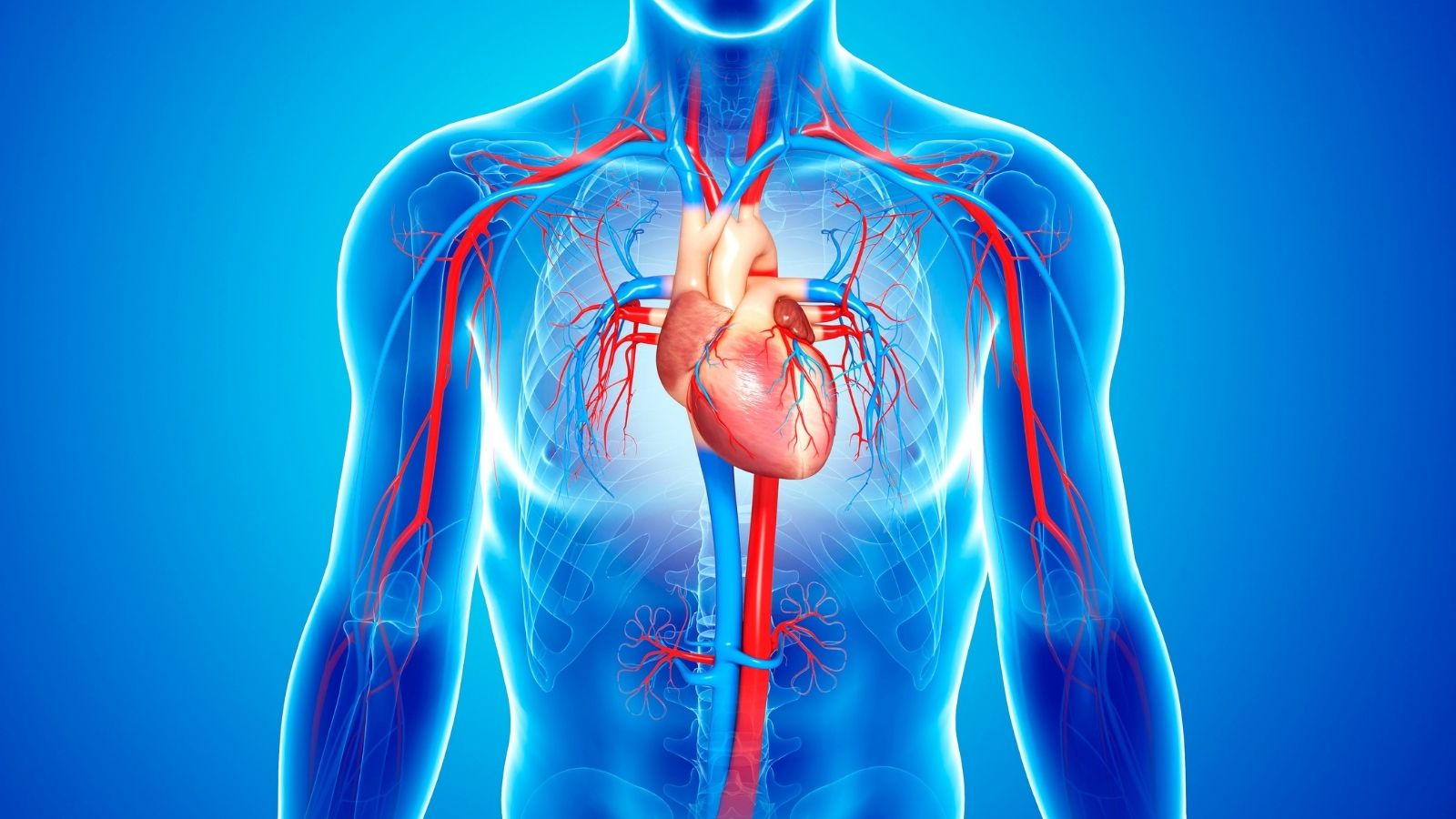
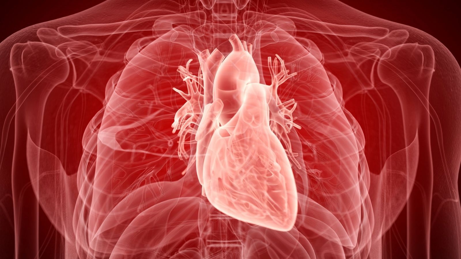
Echocardiography is a non-invasive imaging method that uses ultrasound waves to visualize heart structures and assess function. It is crucial in diagnosing valvular disease, cardiomyopathies, heart failure, and congenital heart defects.
Transthoracic echocardiography is the most commonly used type, providing detailed information about chamber size, wall motion, and valve function. Transesophageal echocardiography offers superior visualization in certain complex conditions.
Doppler echocardiography assesses blood flow and pressure gradients, enabling detection of stenosis, regurgitation, and intracardiac shunts. Stress echocardiography helps evaluate ischemia and guide treatment strategies for coronary artery disease.
The method’s safety, accessibility, and real-time diagnostic capability make it indispensable in clinical practice. Echocardiography guides treatment decisions, monitors therapy effectiveness, and provides valuable prognostic information.
| Definition | A non-invasive diagnostic method that visualizes the structure and function of the heart with ultrasound waves |
| Areas of Use | Valvular heart diseases, heart failure, congenital heart diseases, heart muscle diseases, heart tumors, pericardial diseases, evaluation of heart function |
| Varieties | Transthoracic echocardiography (TTE), transesophageal echocardiography (TEE), stress echocardiography, contrast echocardiography, 3D echocardiography |
| Advantages | Provides painless, rapid, radiation-free, safe, dynamic assessment |
| Preparation Process | Usually requires no special preparation; fasting may be required for TEE |
| Mode of Application | The heart is visualized from various angles with an ultrasound probe by applying gel to the skin, and TEE is performed with a probe through the esophagus |
| Diagnostic Information | Structure and function of the heart valves, size of the heart chambers, contractile force (EF), presence of holes or abnormalities in the heart, fluid accumulation, tumors or clots |
| Complications | Rare in transthoracic ECHO; sore throat in TEE, very rarely risk of esophageal injury |
| Limitations | Obesity, lung disease, chest wall deformities may limit image quality |
What is Echocardiography in Basic Terms?
You can think of echocardiography as an ultrasound of your heart. Just like ultrasound during pregnancy to see how the baby is doing, echocardiography uses sound waves. But this time the focus is not on a baby in the womb, but on the vital organ behind your rib cage. This is why it is called “ultrasound of the heart” and this similarity can help you understand how harmless it actually is.
So how does this technology work? The process is quite simple. It uses a small device that fits in the hand, which we cheaply call a “probe” or “transducer”. This device generates sound waves at a frequency inaudible to the human ear and, when placed on your chest, sends these waves towards your heart. The sound waves hit different structures in your heart, such as muscle tissue, chambers and valves, and are reflected back. We call these reflected waves “echoes”. The device collects these echoes and, with the help of a computer, instantly translates them into a moving, live heart image on a screen. This allows us to monitor in real time every beat of your heart, how it pumps blood and every opening and closing of the valves.
What is one of the best things about this test? Unlike X-rays or CT scans, it contains absolutely no radiation. This makes echocardiography extremely safe. Especially for people whose heart condition needs to be monitored at regular intervals, this test can be repeated over and over again with peace of mind. The test is performed by health professionals specially trained in this field, called “cardiac sonographers”, and the results are carefully reviewed by a cardiologist.
In Which Situations is Echocardiography Ordered?
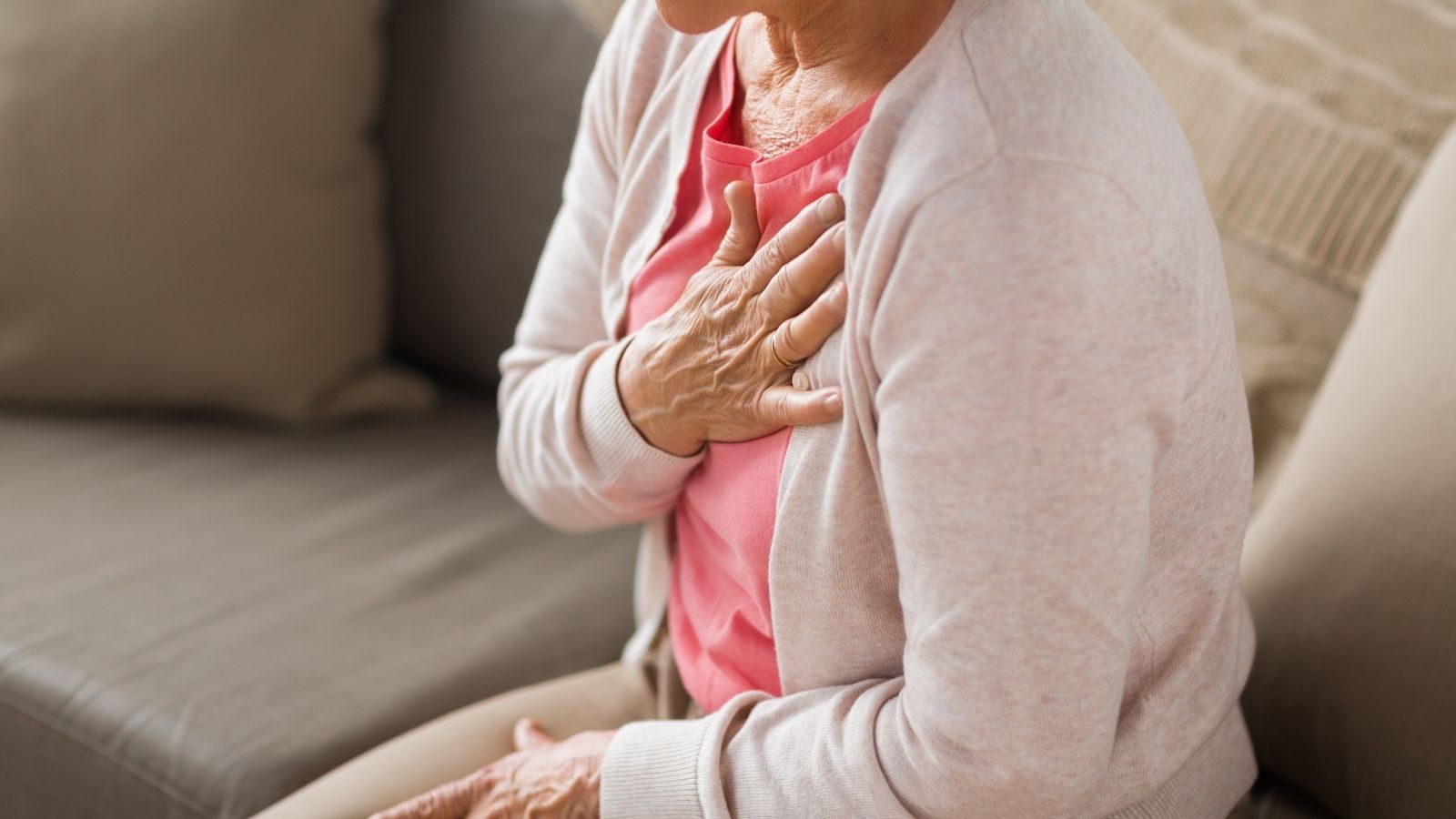
There are many reasons why a doctor may ask you for an echocardiogram. Often it is to find the source of some symptoms you are experiencing or to clarify a condition that was noticed during the examination. Echocardiography works like a detective to clarify a suspicion about the heart. There are some common conditions and symptoms that may cause your doctor to order an echocardiogram.
Your doctor may want to assess the condition of your heart, especially if you experience one or more of the following symptoms
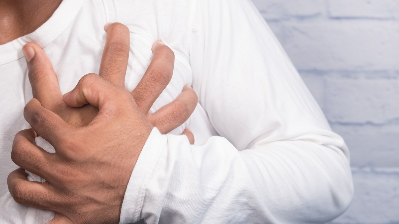
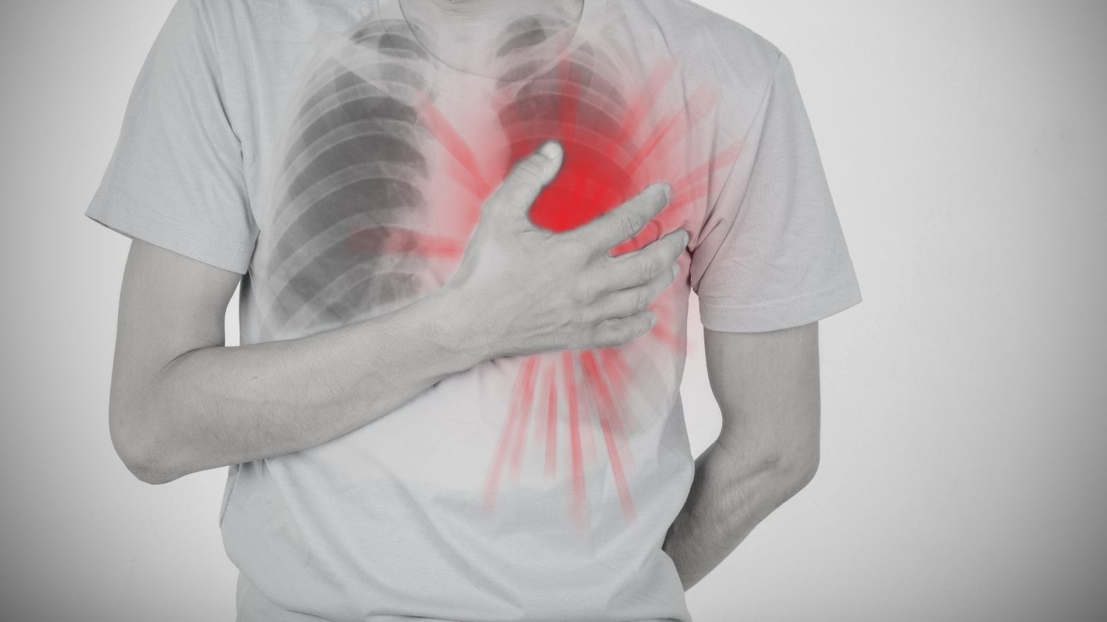
- Pain or tightness in the chest
- Shortness of breath, which is especially pronounced during exertion
- Irregular or rapid heartbeat (palpitations)
- Dizziness or fainting
- Unexplained swelling in the legs, ankles or abdomen (edema)
Sometimes, even if you do not have symptoms, some sounds your doctor hears while listening to your heart with a stethoscope during a routine examination may warrant an echocardiogram:
- Hearing abnormal sounds in the heart called “murmur”
- Detection of suspicious findings on electrocardiogram (ECG) test
- The heart appears larger than normal on chest x-ray
Echocardiography is also an indispensable tool for the diagnosis, follow-up or treatment planning of certain diseases:
- Suspicion and follow-up of heart failure
- Assessing damage to the heart muscle after a heart attack
- Detailed examination of the condition of the heart valves
- Diagnosing congenital heart diseases (such as heart holes)
- Seeing the effect of high blood pressure on the heart
- To assess the general condition of the heart before procedures such as heart surgery or angioplasty
- To check the effectiveness of the heart medication used or the pacemaker implanted
Which Heart Problems Can Be Seen with Echocardiography?
Echocardiography is like a health report card of your heart. It provides your doctor with invaluable information about both the anatomical structure of your heart and how efficiently it works. The ability to detect or exclude many different problems with a single test makes it a cornerstone of cardiology.
Here are some important conditions that can be assessed with echocardiography:
Heart Size and Wall Thickness: It clearly measures whether your heart chambers are wider than normal (dilatation) or whether your heart walls are excessively thickened (hypertrophy), especially due to high blood pressure.
Pumping Power (Ejection Fraction): This is the most important functional measurement of your heart. the “Ejection Fraction” (EF) shows what percentage of the blood in the left ventricle, the main pumping chamber of your heart, is sent to the body each time it contracts. In a healthy heart, this ratio is usually between P-70. A low value may indicate a reduced contractile strength of the heart and a risk of heart failure.
Heart Muscle Damage: Traces of a previous and perhaps unrecognized heart attack can be detected by echocardiography. It can be seen that one area of the heart muscle does not contract as strongly as others (regional wall motion abnormality). This is a sign that the area is not getting enough blood or is permanently damaged.
Problems with Heart Valves: Your heart has four valves that keep the blood flowing in the right direction. Echocardiography examines the structure of these valves in detail. Problems such as calcification, thickening or incomplete closure of the valves may be detected.
Stenosis (Stenosis): The valve fails to open fully, blocking blood flow.
Regurgitation (regurgitation): Incomplete closure of the valve and back leakage of blood.
Blood Clots Blood clots (thrombi) can form inside the heart chambers, especially in certain rhythm disorders (such as atrial fibrillation) or when the heart’s pumping power is severely reduced. Detecting these clots is vital to prevent serious complications such as stroke.
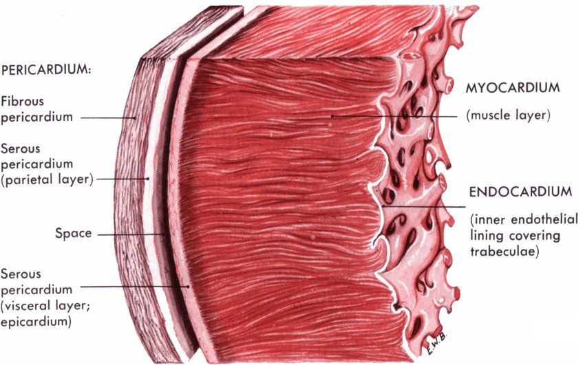
Pericardial Diseases: Conditions such as fluid accumulation in the pericardial membrane (pericardial effusion) or inflammation of the membrane (pericarditis), which surrounds the heart like a sac, can easily be seen.
Congenital Heart Defects: Holes between the chambers of the heart (such as ASD, VSD) or other structural anomalies that can be detected incidentally even in adulthood are diagnosed with this test.
Which Echocardiography Methods Are Available for Heart Evaluation?
There is no single type of echocardiography. There are various methods designed to better assess different aspects of your heart or to find answers to specific questions. Your doctor will choose the one that best suits your situation.
Transthoracic Echocardiography (TTE)
This is the first thing that comes to mind when you think of “ECHO”, the most standardized and most commonly used method. It is performed through a special gel that is applied to the skin over the rib cage. The probe is placed in different positions on the chest to obtain basic and invaluable information about the overall structure and function of your heart. It is usually painless and requires no preparation.
Transesophageal Echocardiography (TEE)
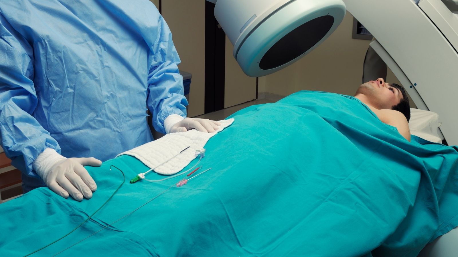
Sometimes standard TTE does not provide a clear enough image. Especially in people who are overweight or have lung disease, the sound waves may have difficulty reaching the heart. We also use TTE when we want to get a much closer look at the back of the heart, valves or possible clots. In this procedure, a thin, flexible tube with a tiny ultrasound probe at the end is inserted through the mouth into the esophagus. Since the esophagus is located directly behind the heart, this method allows us to look into the “backyard” of your heart. This allows for much clearer and more detailed images. Before the procedure, your throat is numbed and a mild sedative is given for your comfort.
Stress Echocardiography
The state of your heart at rest is not always the same as under stress. Some cardiovascular diseases only show symptoms when the heart has to work harder. Stress echocardiography is performed to reveal this. The stages of the test are as follows.
First, an ECHO is performed at rest.
You will then be asked to walk on a treadmill or cycle on a stationary bike. If you are unable to exercise, a medicine is given intravenously that speeds up your heart as if you were exercising.
As soon as the effect of the exercise or medication is maximized, a second ECHO is performed immediately.
The two ECHO recordings are compared to see how your heart reacts to stress and whether there is enough blood supply to the heart muscle.
Doppler Echocardiography
This technique is an integral part of the standard ECHO examination. It shows not only the structure of the heart but also the movement of blood inside it. The Doppler technique measures the speed and direction of blood flow. With this information we can assess the following:
- The speed of blood passing through the heart valves
- Whether there is any leakage in the covers
- Pressures between the heart chambers
- Blood flow pattern inside the heart
On the screen, we usually see blood flow color-coded in red and blue. This allows us to recognize abnormal currents immediately.
Three Dimensional (3D) Echocardiography
This is one of the most advanced versions of echocardiography. It goes beyond traditional two-dimensional (2D) imaging to create a live, three-dimensional model of your heart. It provides invaluable information, especially for a surgeon planning to repair the complex structure of the heart valves or to measure the volume of the heart chambers in the most precise way.
How to prepare for an echocardiography test?
Preparation for an echocardiography test varies depending on which type of ECHO you will have. It is therefore very important that you follow the instructions given to you by your doctor or the center where the test will be performed.
Preparation for standard (Transthoracic) ECHO:
This most common test usually requires no special preparation. One of the most frequently asked questions is whether the heart echo is performed on an empty stomach. You do not need to be hungry for a standard echo. You can eat normally before the test, drink water and take your daily medication unless your doctor tells you otherwise. You only need to wear comfortable clothes.
Preparation for transesophageal echocardiography (TEE):
This test requires more serious preparation as it involves inserting a tube into the esophagus.
Fasting: You are asked not to eat or drink anything at least 6, preferably 8 hours before the procedure. Having a completely empty stomach eliminates the risk of nausea and vomiting during the procedure.
Medicines Be sure to inform your doctor about any medicines you are taking, especially blood thinners or diabetes medicines. You will be given specific instructions about when to take or stop taking which medicine.
Accompaniment Since you will be sedated during the procedure, you may feel dizzy afterwards. For this reason, driving is strictly prohibited on the day of the procedure and you must have someone with you who can drive you home.
Preparation for stress echo:
As this test involves physical exertion, some preparation is important.
Eating and drinking: You can eat something light about 3-4 hours before the test. On the day of the test, however, you are asked to avoid caffeinated drinks such as coffee, tea, cola and cigarettes because they can affect your heart rate.
Clothing: It is important to wear comfortable, exercise-friendly clothes (such as sweatpants) and sneakers.
Medicines: Your doctor may ask you to stop taking certain heart medications (especially beta blockers that slow the heart rate) a day or two before the test, which may affect the accuracy of the test. Never stop taking any medication without consulting your doctor.
What Happens During the Echocardiography Procedure?
Knowing what to expect at the time of the procedure can help reduce your anxiety. How to take a standard echo? The process is as follows step by step:
When you are taken into the room where the test will be performed, you will be asked to undress from the waist up and put on a patient gown that will be given to you. The procedure is exactly the same for female patients. You will then be asked to lie on an examination table, usually on your left side. This position improves image quality by bringing your heart closer to the chest wall.
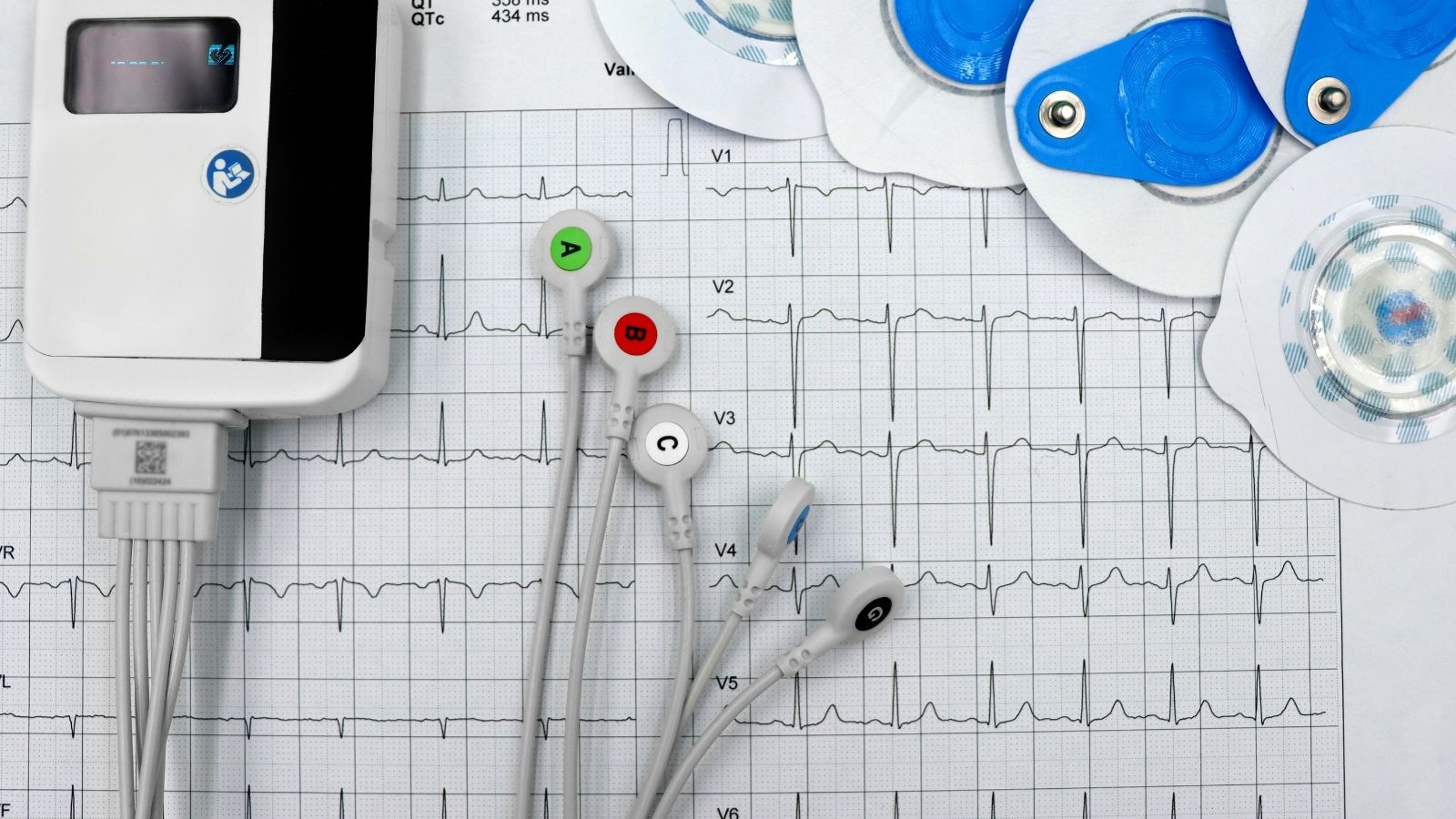
The technician sticks several small, sticky strips (ECG electrodes) to your chest to monitor your heart rhythm throughout the test. These are completely harmless. He or she will then apply a warm, slippery gel to specific areas of your chest to ensure that there is no air between your skin and the ultrasound probe and to help the sound waves transmit better. This gel is water-based and does not stain.
The technician then begins to record images of your heart by moving the ultrasound probe over the gel areas and occasionally pressing gently to get a clearer image. This pressure can sometimes feel slightly uncomfortable, but usually does not cause pain. During the procedure, you may be asked to hold your breath for a short time or to assume a different position. These small maneuvers allow us to see parts of the heart better by pulling organs such as the lungs out of the way.
So, how long does a standard echocardiogram take? The procedure is usually completed in 30 to 60 minutes. When the procedure is finished, the gel is removed and you can immediately return to your normal life.
It is a painless and radiation-free imaging method that uses sound waves to visualize the structure, movement and blood flow of the heart. It is used to evaluate many conditions such as valvular heart disease, heart failure, congenital heart disease, thickening of the heart muscle and the presence of clots. It provides information about the size of the heart chambers, the strength of contraction, valve function, the presence of fluid and the condition of the great vessels. No, it does not contain radiation. Since sound waves are used, it can be safely applied in pregnant women and children. Standard transthoracic echocardiography usually takes 15-30 minutes. Gel is applied to the chest wall and the heart is visualized with the probe. The patient lies supine or on the left side. The patient should remain calm and follow the technician’s instructions. No need to starve. There are types such as transthoracic (TTE), transesophageal (TEE), stress echocardiography and fetal echocardiography. It is a method that allows a clearer visualization of the posterior structures of the heart with a special probe inserted into the esophagus. By accelerating the heart with exercise or medication, cardiac performance during exertion is evaluated. It is used to detect coronary artery disease. It is applied between 18-24 weeks of pregnancy to evaluate the baby’s heart structure. Most of the time, the cardiologist evaluates the imaging immediately and a report is generated in a short time. Yes, if the contractile strength of the heart muscle decreases after a heart attack, it can be detected by ECHO. If there is diagnosed heart disease, it is done at regular intervals. If there is no risk, it is only taken in suspicious cases. Frequently Asked Questions
What is Echocardiography (ECHO)?
For what purposes is ECHO performed?
What information is obtained with ECHO?
Is ECHO harmful?
How long does ECHO take?
How to take an ECHO?
What to look out for during ECHO?
How many types of ECHO are there?
What is Transesophageal ECHO?
What is stress echocardiography for?
When is Fetal ECHO performed?
[/accordion-item] Are ECHO results available immediately?
Can ECHO detect a heart attack?
How often should ECHO be performed?

Prof. Dr. Yavuz Beşoğul graduated from Erciyes University Faculty of Medicine in 1989 and completed his specialization in Cardiovascular Surgery in 1996. Between 1997 and 2012, he served at Eskişehir Osmangazi University Faculty of Medicine as Assistant Professor, Associate Professor, and Professor, respectively. Prof. Dr. Beşoğul, one of the pioneers of minimally invasive cardiovascular surgery in Türkiye, has specialized in closed-heart surgeries, underarm heart valve surgery, beating-heart bypass, and peripheral vascular surgery. He worked at Florence Nightingale Kızıltoprak Hospital between 2012–2014, Medicana Çamlıca Hospital between 2014–2017, and İstinye University (Medical Park) Hospital between 2017–2023. With over 100 publications and one book chapter, Prof. Dr. Beşoğul has contributed significantly to the medical literature and is known for his minimally invasive approaches that prioritize patient safety and rapid recovery.