Angiography is a diagnostic imaging procedure that visualizes blood vessels using contrast dye and X-ray technology. It is primarily used to detect blockages or narrowing in coronary arteries.
Indications for angiography include chest pain, suspected coronary artery disease, abnormal stress test results, or acute heart attack. It provides detailed information about blood flow and vessel integrity.
The procedure involves inserting a catheter, usually through the femoral or radial artery, and injecting contrast dye. Real-time imaging shows the vascular structure and any obstructions present.
Treatment decisions such as angioplasty or stent placement are often made based on angiography findings. The procedure is generally safe but carries small risks of bleeding, arrhythmia, or allergic reactions.
| Medical Name | Angiography (Anjio) |
| Frequent Use Areas | – Diagnosis of coronary artery disease- Diagnosis of peripheral artery and vein diseases- Aneurysm, vascular occlusion, vascular stenosis research |
| Causes | – Detailed visualization and evaluation of the vascular structure – Suspicion of obstruction, narrowing or aneurysm |
| Risk Factors | – Allergy to contrast media – Impaired renal function – History of bleeding and vascular disease – Advanced age |
| Complications | – Bleeding- Infection- Allergic reaction due to contrast media- Vascular injury- Impaired renal function |
| Diagnostic Methods | – Clinical examination- ECG- Blood tests- CT/MR or ultrasound may be required for preliminary evaluation |
| Treatment Methods | – Angiography is a diagnostic method; if necessary, balloons, stents or other interventional treatments can be performed during the procedure |
| Prevention Methods | – Evaluation of renal function before the use of contrast media- Taking necessary precautions in patients with a history of allergy |
What is Angiography?
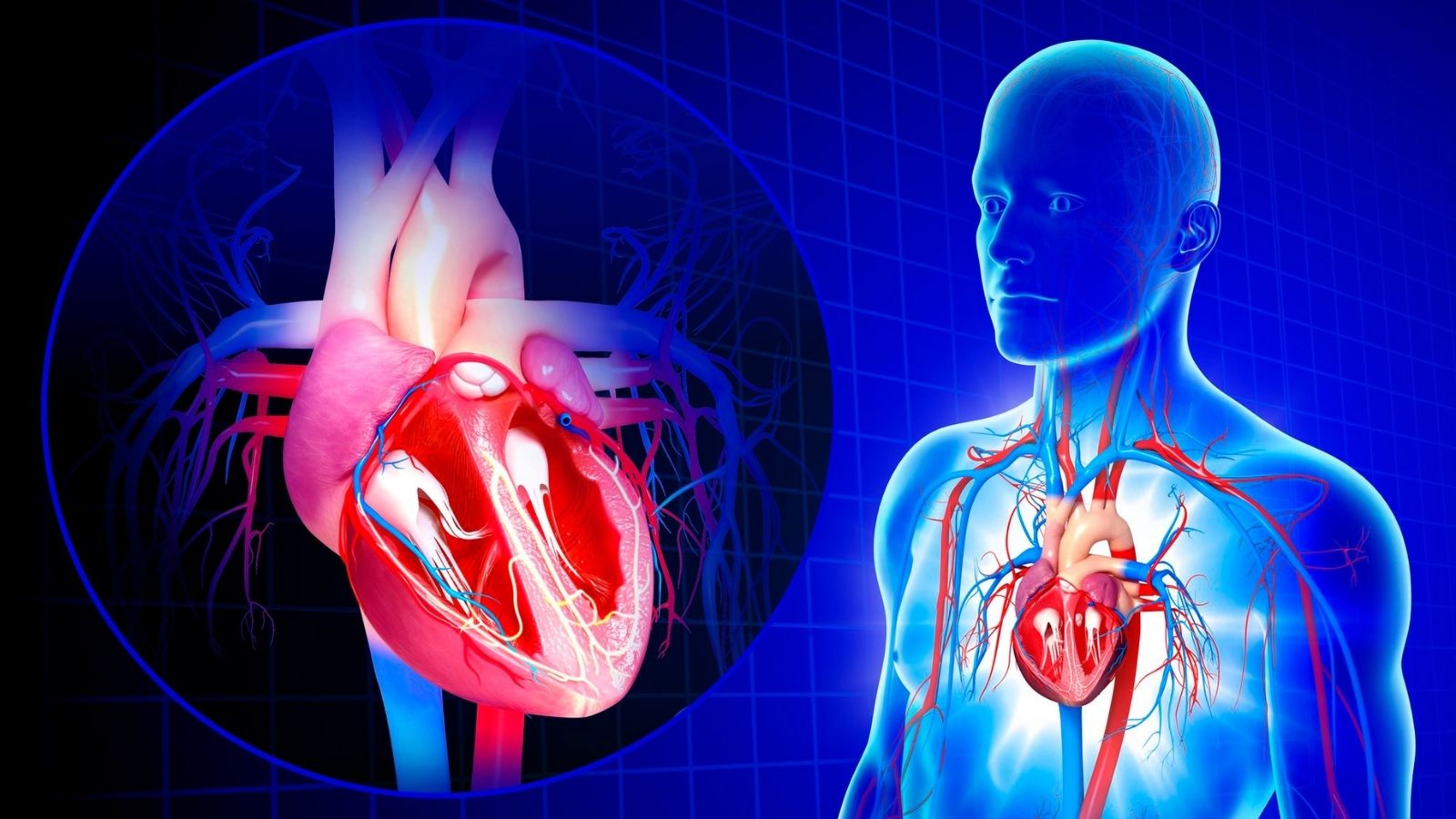
In its simplest form, angiography is an advanced X-ray technique that creates a kind of “road map” of the vascular network in our body. The arteries, veins and chambers of the heart are invisible on a normal X-ray. Angiography is the art of making these invisible vessels visible. To do this, we use a special dye called “contrast dye”. When we introduce this dye into the blood vessels, the vessels become brightly visible under X-rays. This allows us to clearly see how the blood flows through the vessels and whether there is any narrowing, blockage, bubbling (aneurysm) or other structural defects in the vessel walls.
The images obtained by this procedure are called “angiograms”. These images are not just like a photo frame; they are like a live video recording showing the inside of the vessels. Thanks to this technology, we can examine the vascular system in almost every part of the body in detail, from the coronary arteries that feed the heart to the carotid arteries that carry blood to the brain, from the kidney veins to the leg veins.
Why is Cardiac Angiography Necessary and What Diseases Does It Diagnose?
Angiography is considered the “gold standard” in the diagnosis of cardiovascular diseases. So why does a doctor recommend an angiogram to a patient? There are several main reasons for this:
The most common cause is suspected Coronary Artery Disease. Over time, cholesterol and lime build up on the walls of the thin vessels that feed our heart, called coronary arteries, and form plaques. These plaques narrow the arteries and prevent the heart from being adequately supplied with blood. Angiography is the most precise method that shows us where these stenoses are, how many they are and how serious they are.
Our reasons for seeking angiography may include
- Coronary artery disease (CAD)
- Chest pain (angina pectoris)
- Shortness of breath
- Quick fatigue
- Heart attack (acute myocardial infarction)
- Abnormal stress test or other heart tests
- Heart valve disease
- Congenital heart anomalies
- Aortic vessel enlargement (aneurysm) or rupture (dissection)
- Narrowing of the leg arteries (peripheral arterial disease)
- Narrowing of the kidney vessels (renovascular hypertension)
- Blockage or bubbles in the brain vessels
This method is not only a diagnostic tool; it is also the basis for treatment planning. For example, the vascular structure in the angiogram is the most important guide for us when deciding whether bypass surgery or stenting is more appropriate for a patient.
What Should Be Done Before Angiography?
When the decision for angioplasty is made, the process actually starts before the day of the procedure. This preparation period is very important for both the safety of the procedure and your comfort. Here are some key points to consider before an angioplasty.
Hunger: Your stomach must be empty before the procedure. For this reason, you are usually asked not to eat or drink anything, including water, from 6 to 8 hours before the procedure, i.e. after midnight the night before.
Medicines It is vital that you fully inform your doctor of all medications you are taking (prescription, over-the-counter, vitamins, herbal supplements). In particular, your doctor will give you specific instructions about blood thinners (such as Coumadin, Plavix, Xarelto, etc.) and diabetes medications (such as Metformin). These medicines may need to be stopped or the dosage adjusted a few days before the procedure.
Allergies If you have a known allergy to iodized dyes, seafood, latex or any medication, you should tell your healthcare team. If necessary, you may be given protective medication before the procedure to prevent an allergic reaction.
Kidney Functions: The dye used in angiography is excreted through the kidneys. Therefore, your doctor will order a simple blood test to check your kidney function before the procedure. If your kidneys are sensitive, additional IV therapy may be administered before and after the procedure to protect the kidneys.
Accompanying person: During the procedure you will be given a mild sedative to help you relax. As the effect of this medication may last for some time, it is not safe for you to drive after the procedure. You should therefore arrange in advance for a relative to take you home from the hospital.
How is the Angioplasty Procedure Performed?
Let’s talk step by step about what to expect when you arrive at the hospital on the day of the angioplasty. Knowing this process will help reduce your worries.
First, you will be taken to a private room and asked to put on a comfortable patient gown. The nurses will check your vital signs, such as blood pressure and pulse rate, and start an IV in your arm. This intravenous line is used to give you the necessary medicines and fluids during the procedure.
The angio lab where the procedure will take place is a special room equipped with technological devices. Once in the room, you lie on the procedure table and ECG electrodes are attached to your chest to continuously monitor your heart rhythm.
Angioplasty is usually performed at one of two main sites:
- The artery in the wrist (radial artery)
- The artery in the groin (Femoral artery)
The doctor decides which area is more suitable for you based on your vascular structure and the nature of the procedure. Today, the wrist is generally preferred because of its comfort and safety advantages.
The selected area (wrist or groin) is cleaned with an antiseptic solution and then numbed with a local anesthetic. This is similar to the way your dentist numbs your tooth; you will only feel a slight burning sensation at the insertion of the first needle, after which the area is completely numb.
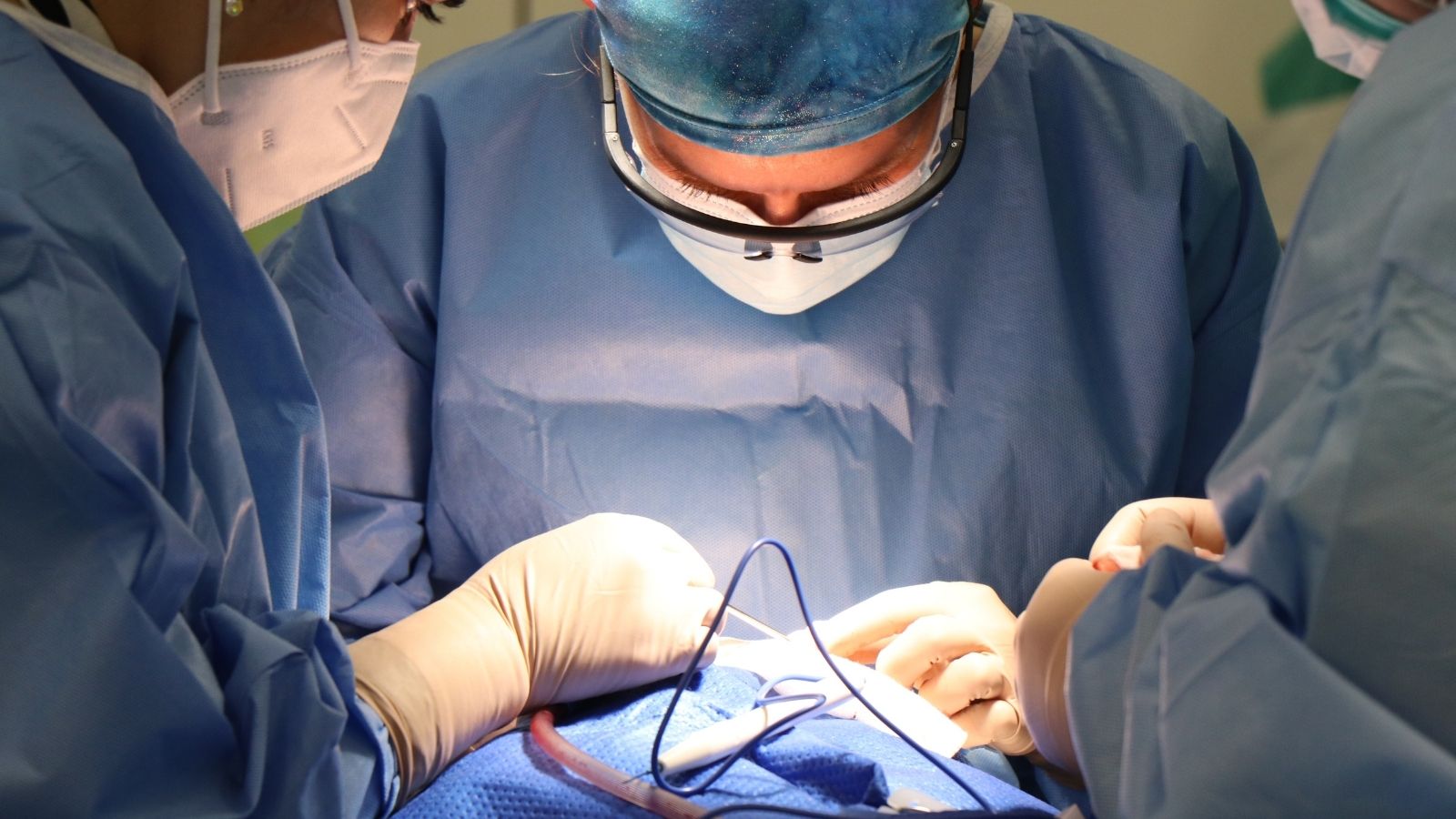
A thin, plastic tube called a “sheath” is inserted into the artery at the numbed site. This sheath acts as a door to the artery throughout the procedure. The whole procedure is done through this sheath. A very thin, flexible and long tube called a “catheter” is then inserted through this sheath. Watching through a live X-ray screen called fluoroscopy, the doctor gently guides this catheter through your veins and into your heart. You will feel absolutely no pain because there are no pain nerves inside the veins. There may only be a slight feeling of pressure.
When the catheter is inserted into the mouth of the coronary arteries that supply your heart, a dye (contrast dye) is injected through it. At the moment the dye is injected, you may feel a flush of warmth or urge to urinate, which lasts for a few seconds. This feeling is completely normal and temporary and you do not need to worry. While the dye is filling the vessels, the X-ray machine records angiograms (angiograms) in series from different angles. You may be asked to hold your breath briefly or remain still to get clear images.
If it is just a diagnostic angiogram, the procedure is usually completed in 15-30 minutes. After the images are taken, the catheter and sheath are removed, pressure is applied to the entry site to stop bleeding and a tight bandage is applied.
How is Inguinal Angioplasty Performed and When Is It Preferred?
Inguinal angiography, or femoral approach, is the more traditional method of angiography and is still indispensable in certain situations. So, how is an angiography through the groin performed and why is this route sometimes preferred over the wrist? The process is very similar to wrist angiography. The stages of preparation and admission to the laboratory are the same. The main difference is that the entry point is in the groin. After the groin area has been cleaned and numbed, a needle is inserted into the femoral artery (the main artery in the groin) and the sheath is placed there. The catheter is guided through this sheath into the aorta, the main artery of the body, and from there into the vessels of the heart.
There are some special cases where angioplasty through the groin is preferred:
- The veins in the wrist are too thin, tortuous or blocked.
- There has been a previous unsuccessful attempt on the wrist.
- Complex procedures that require the use of much thicker or specialized medical devices (e.g. large stents, heart pumps).
- Patients who have had some bypass surgery (especially bypasses using leg veins).
- Fistula for dialysis in the patient’s arm veins.
Because the femoral artery is a much larger vessel than the radial artery, it offers the doctor a larger working area and more support, especially in difficult and complex cases. However, the healing process is a little more laborious than for the wrist.
Are There Factors Affecting Angiography Image Quality?
Yes, the quality of the angiogram image we obtain is critical for us to make an accurate diagnosis. Several factors affect the clarity and interpretability of the image:
Patient Movement: It is very important for the patient to remain still during the procedure, especially while the dye is being administered, and to hold their breath when instructed to do so. Movement can cause images to blur.
Body Structure: In overweight or muscular patients, it may be more difficult for X-rays to penetrate the tissue, which may slightly reduce image quality.
Vessel Anatomy: Very tortuous or overlapping vessels can make it difficult to visualize the stenosis clearly. This is why we rotate the X-ray machine at different angles (right anterior oblique, left anterior oblique, etc.) to get the best view of each vessel segment.
Calcification: Dense calcification in the vessel wall can block X-rays and over- or underestimate the degree of stenosis. In such cases, we may resort to additional imaging modalities such as intravenous ultrasound (IVUS).
Equipment Used: Modern angiography devices can produce much higher resolution images with a lower radiation dose. The quality and technology of the device directly affect the results.
What are the Benefits and Harms of Angioplasty?
Like any medical procedure, angiography has potential benefits and risks. The important thing is to strike the right balance between the two.
Benefits of Angiography:
The greatest benefit of angiography is its unerring accuracy in diagnosing vascular diseases. By showing us directly inside the vessel, it tells us exactly what the problem is and how serious it is. But the truly revolutionary benefit is the ability to “see and treat”.
What does this mean? If during the angiogram we detect a stenosis that severely obstructs blood flow, we can proceed with treatment in the same session without terminating the procedure.
Balloon Angioplasty: We access the area of stenosis with a balloon catheter, inflate the balloon, crush the plaque and open the vessel.
Stent Placement: To prevent the vessel from narrowing again, we insert a mesh-like tube made of metal, called a “stent”, into the opened area. This stent acts as a scaffold for the vessel, keeping it open.
This approach is especially life-saving in emergencies such as heart attacks. In a heart attack, we say “time is muscle” because the sooner the blocked vessel is opened, the more heart muscle tissue can be saved. Angiography and immediate stenting minimize or completely prevent permanent damage to the heart.
Potential Harms (Risks) of Angion:
Although angiography is an extremely safe procedure when performed in experienced hands, it carries some risks as it is an invasive (interventional) procedure. It is important to know these risks and to talk about them transparently.
The most common side effects, which are usually mild, are
- Bruising or bleeding (hematoma) at the procedure site.
- Mild pain or tenderness at the insertion site.
- Transient hot flushes due to contrast medium.
More rare but potentially serious risks include the following:
- Allergic reactions to the contrast medium.
- Temporary impairment of kidney function (especially in patients with pre-existing kidney disease).
- Damage to the access vessel (pseudoaneurysm, fistula).
- Heart attack.
- Stroke (paralysis).
- Blood clot formation.
Is Angioplasty Dangerous and Is There a Risk of Death Due to the Procedure?
This is the question that patients are most curious and concerned about. To put it bluntly, every surgical or interventional procedure has a risk. However, thanks to modern medical technology, advanced materials and experienced teams, angiography has become an extremely safe procedure.
Statistically, the risk of serious complications (e.g. heart attack, stroke) from a diagnostic angiography is very low, usually less than 1%. The risk of death from angioplasty is much rarer and is expressed in rates of less than one in a thousand (about 0.08%).
This is where the risk-benefit balance comes into play. If a patient is at high risk of having a heart attack, or if their current symptoms seriously impair their quality of life, the diagnostic and therapeutic benefit of angiography far outweighs these very low statistical risks. Your doctor will carry out this risk-benefit analysis with you, taking into account your personal health condition, age and comorbidities.
What Should Be Considered After Angioplasty?
The procedure is over, now what? Recovery after angioplasty varies slightly depending on where the procedure was performed.
If the procedure is performed on the wrist: You can usually stand up, walk and take care of your basic needs shortly after the procedure. A special bandage on your wrist is removed after a few hours. The healing process is quite comfortable.
If the procedure is done in the groin: Because the vein in the groin is larger, you will need to lie on your back and without bending your leg for several hours (usually 4-6 hours) to prevent bleeding. At the end of this time, you can stand up under the control of the nurse.
There are some general rules to follow after returning home:
Drink plenty of fluids: Drink plenty of water on the first day to help your body easily remove the dye through the kidneys.
Rest: Avoid strenuous activities, heavy lifting and sudden movements for the first 24-48 hours after the procedure.
Entry Site Care: Keep the treated area clean and dry. You can remove the bandage and shower according to the instructions given to you. A slight bruising or a small, firm swelling in the area is normal and disappears over time.
Medicines It is very important that you take the medicines (especially blood thinners) prescribed by your doctor regularly.
If you experience any of the following situations, you should contact your doctor or the nearest emergency room immediately:
- Active bleeding at the site of the procedure that does not stop with pressure.
- Increasing, severe pain, swelling or redness at the insertion site.
- Coldness, pallor or numbness in the treated arm or leg.
- Severe chest pain or shortness of breath.
- Sudden dizziness or fainting.
How many times can a person have an angioplasty?
This is also a frequently asked question. There is no set number or “quota” for angiography. The number of times a person can have an angioplasty in a lifetime depends entirely on medical requirements.
Since angiography is a procedure involving radiation, it is not repeated arbitrarily or unnecessarily. However, if a patient’s medical condition requires it, angiography may be performed repeatedly. For example:
- To evaluate new complaints of a patient who had a stent implanted years ago.
- To check the condition of the bypass vessels of a patient who has had bypass surgery years later.
- When new problems arise in different vascular regions (e.g. the heart first, then the leg veins years later).
Each angioplasty decision is based on a risk-benefit analysis, taking into account the patient’s current condition, complaints and previous test results. It is important that the benefits of the procedure outweigh the potential risks. Since radiation doses are minimized with modern devices, it is safe to repeat the procedure if necessary.

Prof. Dr. Yavuz Beşoğul graduated from Erciyes University Faculty of Medicine in 1989 and completed his specialization in Cardiovascular Surgery in 1996. Between 1997 and 2012, he served at Eskişehir Osmangazi University Faculty of Medicine as Assistant Professor, Associate Professor, and Professor, respectively. Prof. Dr. Beşoğul, one of the pioneers of minimally invasive cardiovascular surgery in Türkiye, has specialized in closed-heart surgeries, underarm heart valve surgery, beating-heart bypass, and peripheral vascular surgery. He worked at Florence Nightingale Kızıltoprak Hospital between 2012–2014, Medicana Çamlıca Hospital between 2014–2017, and İstinye University (Medical Park) Hospital between 2017–2023. With over 100 publications and one book chapter, Prof. Dr. Beşoğul has contributed significantly to the medical literature and is known for his minimally invasive approaches that prioritize patient safety and rapid recovery.