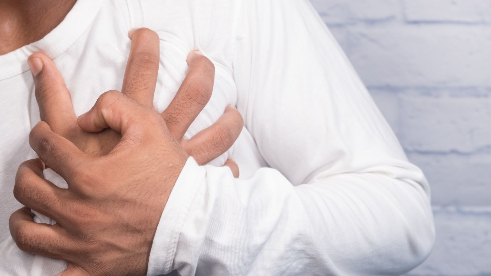
Heart transplantation is a surgical procedure performed in patients with end-stage heart failure when medical or device-based treatments are no longer sufficient. It involves replacing the diseased heart with a healthy donor heart.
Candidate selection for heart transplantation is critical, requiring thorough evaluation of organ function, comorbidities, and overall health status. Patients must meet strict criteria to ensure success and minimize postoperative risks.
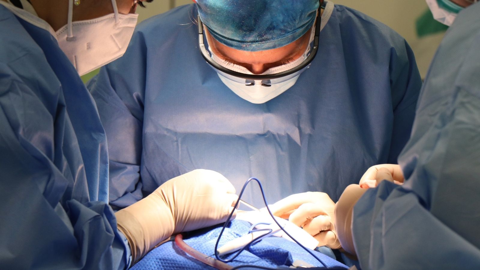
The surgery is performed under general anesthesia, involving removal of the failing heart and connection of the donor heart to major vessels. Postoperative intensive care monitoring is essential to detect complications early.
Long-term success depends on immunosuppressive therapy, lifestyle adjustments, and regular follow-up. Despite risks such as organ rejection and infection, heart transplantation significantly improves survival and quality of life in selected patients.
| Medical Name | Cardiac Transplantation (Heart Transplantation) |
| Common Uses | – End-stage heart failure – Severe cardiomyopathy – Advanced heart muscle diseases – Patients who have not responded to other therapies |
| Causes | – Advanced heart failure – Ischemic heart disease – Dilated cardiomyopathy – Congenital heart disease (congenital) |
| Risk Factors | – Long-term heart disease – Underlying chronic diseases – Previous heart surgery |
| Complications | – Acute or chronic tissue rejection – Risk of infection – Side effects of immunosuppressive drugs – Development of coronary artery disease |
| Diagnostic Methods | – Echocardiography- ECG- Blood tests- Tissue and compatibility tests- Cardiac MRI or CT |
| Treatment Methods | – Surgical transplantation of a suitable donor heart – Lifelong immunosuppressive (immunosuppressive) drug therapy |
| Prevention Methods | – Prevention of infections – Regular medical follow-up – Regular use of immunosuppressive drugs – Healthy lifestyle |
What are the Requirements for a Heart Transplant and Who Can Be a Candidate?
The decision to have a heart transplant is one of the most important milestones in a person’s life and is never taken lightly. To embark on this path, a patient must be at a point we call “end-stage heart failure”. This means that your heart is no longer able to meet your body’s basic needs despite the most advanced drug therapies, pacemakers or other supportive devices. To find out if this critical threshold has been crossed, a series of objective assessments are performed. These assessments guide both physicians and patients by drawing a kind of road map.
In this process, there are key indicators that determine whether a patient is a transplant candidate. These are scientific data that give a clear picture of the general condition of the patient.
Some of the key clinical signs used in the assessment are:
Measurement of Exertional Capacity (CPET): This test is one of the most important measurements to show how enduring a patient is and how efficiently their body uses oxygen. If the peak oxygen consumption (VO2 max) drops below a certain level as a result of the test, this is the clearest evidence that the heart is no longer able to carry the burden of daily life.
Measurement of Intracardiac Pressures (Cardiac Catheterization): During this procedure, a thin tube (catheter) is inserted into the heart to directly measure pressures and the amount of blood pumped by the heart. If the heart’s pumping power (cardiac index) has fallen too low and the filling pressures inside the heart have risen too high, this is a sign that the heart is hemodynamically failing, i.e. in serious danger.
Course of Clinical Progress: Sometimes more than numbers are needed. The general condition of the patient is also a very important indicator. A patient is constantly hospitalized for heart failure, becoming dependent on intravenous heart-enhancing drugs to sustain life, life-threatening arrhythmias that do not go away despite all treatments, or the gradual deterioration of other organs such as the kidneys and liver due to the weakening of the heart, are alarm bells that indicate that transplantation should now be seriously considered.
Risk Scoring Models: Using advanced medical calculation methods, many parameters such as the patient’s age, blood values, heart function, etc. are combined. These models estimate the probability that the patient will survive one year in their current condition. If this probability falls below a certain percentage, the risk of the disease itself is higher than the risk of heart transplant surgery and this supports the decision to transplant.
What are the Obstacles to Heart Transplantation and How Does the “Artificial Heart” Offer a Solution?
Heart transplantation is not a suitable treatment for every patient. Some health problems can unacceptably increase the risks of this major surgery and make the transplant unsuccessful. It is possible to divide these obstacles into “definite” and “relative”. In the presence of absolute barriers, no transport can be carried out, while temporary barriers can be corrected or managed over time.
Here are some of the conditions that absolutely prevent a heart transplant:
- Cancer that is active and at risk of spreading
- A serious uncontrolled infection in the body
- Severe and irreversible renal or hepatic failure
- A proven history of persistent nonadherence to treatment
- Active alcohol or substance dependence
- Inadequate family or social support
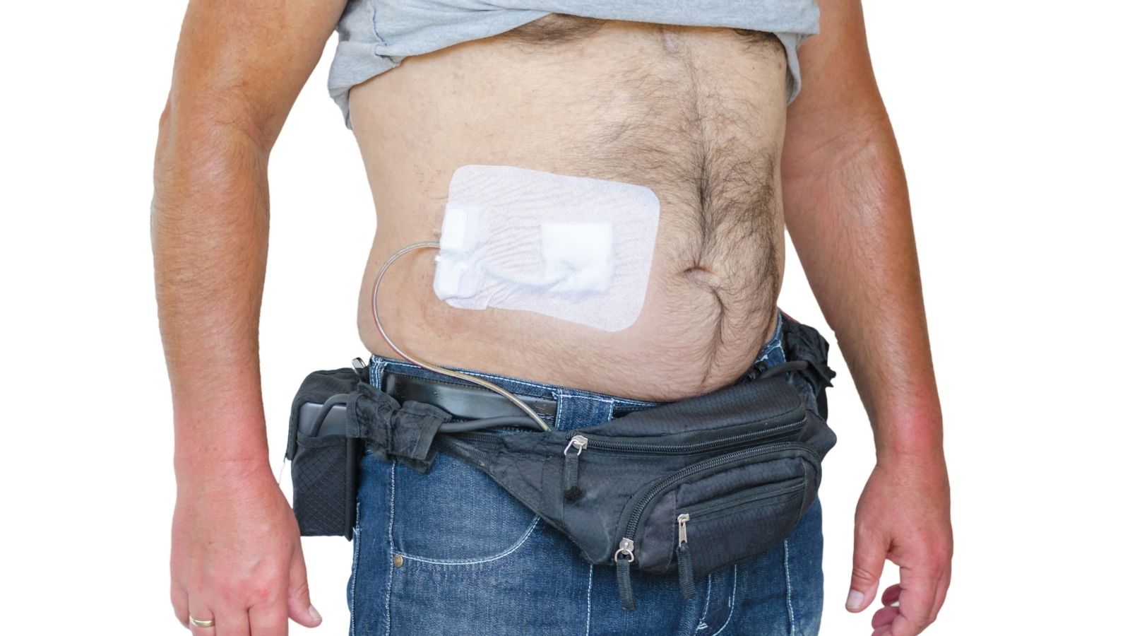
Fortunately, thanks to advances in medicine, some obstacles can now be overcome. There is now a glimmer of hope for many patients who used to be told “they cannot have a transplant”. This is where Ventricular Assist Devices (VAD), also known as artificial hearts, come into play. These devices are sophisticated pumps that take over the pumping role of the heart and can act as a “bridge” to remove some of the more substantial obstacles.
Some commonly recognized barriers that can be overcome with VAD:
High Lung Pressure (Pulmonary Hypertension): This is perhaps the most commonly encountered relative barrier. If the pressure in the lung vessels is too high, the right side of the newly implanted heart may not be able to withstand the pressure and may fail. When a patient is implanted with a VAD, the heart’s load is reduced and over time this high pressure in the pulmonary vessels can decrease to normal levels. The patient thus becomes a safe candidate for transplantation.
Overweight (Obesity): Obesity increases the risk of surgery and complications such as wound infection. A patient under VAD support can get help from a dietitian to lose weight in the process, or even undergo procedures such as gastric reduction surgery to reach the ideal weight for transplantation.
Excessive Weakness and Muscle Loss (KaÅeksi): In the final stages of heart failure, the body can waste away. The insertion of a VAD allows the patient to eat and laugh normally again. With physical therapy and nutritional support, the patient regains the strength to withstand major surgery.
Controllable Kidney Failure: Heart failure can also fatigue the kidneys. When blood circulation improves with VAD, kidney function often improves as well, and the patient may become eligible for a heart transplant alone, without the need for a double organ transplant.
This strategy of “candidate screening” has opened the door of hope to more patients as doctors navigate the heart transplant process. Many obstacles are no longer seen as insurmountable walls, but as challenges that can be jumped over or overcome.
How to Find a Suitable Heart and What is the Process for Patients Waiting for a Heart Transplant?
When a transplant decision is made for a patient, their name is registered on the Ministry of Health’s National Organ Transplant Waiting List. From then on, a patient and transplant team wait patiently. The process is based entirely on objective criteria such as medical urgency, blood type and tissue compatibility.
Once a suitable heart is found, the race against time begins. The lifespan of a heart is very limited after it is taken from the donor. Advanced technologies are used to protect this precious organ and deliver it to the recipient in the healthiest way possible.
Types of donors according to organ source are as follows:
Brain Dead Donors (DBD): Traditionally, most heart transplants are performed from donors whose brain function has completely and irreversibly stopped, but whose heart is still beating.
Post-Circulatory Donors (DCD): This is a new method that has revolutionized the field of heart transplantation in recent years. In patients with severe brain damage but not legally diagnosed as brain dead, life support is terminated with family consent. Shortly after the circulation stops, the heart is removed using special techniques.
Extended Criteria Donors (ECD): Due to the scarcity of organ donation, some formerly unsuitable donors can now be accepted after careful evaluation. For example, the hearts of donors who are slightly older or have some chronic diseases under control can be a chance of life for critically ill recipients by balancing the risks and benefits.
The most innovative technology used to preserve and transport the heart is the “Normothermic Ex-Vivo Perfusion” system. This system, popularly known as “Heart in a Box”, is a machine that keeps the heart at body temperature, feeds it with blood and keeps it functioning, rather than keeping it in a container of ice.
The main advantages of this technology:
- It preserves the vitality of the heart for much longer.
- Allows the delivery of organs from long distances.
- allows for pre-transplant testing of the performance of a heart that is considered “on the edge”.
- It has a positive effect on the postoperative heart transplant success rate by minimizing damage to the heart.
So, how is a heart transplant performed and what are the stages of the surgery?
The answer to the question of how a heart transplant is performed lies in a complex surgical process that requires great care, experience and teamwork. As soon as the transplant team receives news of a suitable heart, the recipient patient is immediately prepared for surgery.
The surgical process proceeds step by step. Each stage is critical to the success of the next.
Preparation and Cardiothoracic Machine: The surgery begins with a midline opening of the chest cage. To maintain the body’s circulation, the patient is connected to a cardiopulmonary machine that oxygenates the blood and pumps it back into the body. This machine temporarily takes over the work of the heart and lungs, which will be stopped during surgery.
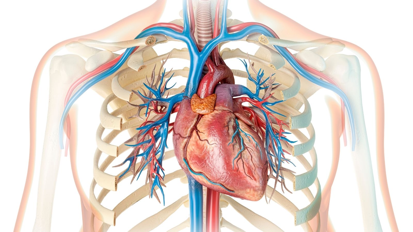
Removal of the diseased heart: After the heart-lung machine is activated, the surgeon carefully removes the patient’s tired and diseased heart. During this procedure, the basic structures where the new heart will be sutured are preserved, namely the posterior wall of the left auricle, where the vessels that bring clean blood from the lungs open, and the ends of the main veins that collect dirty blood from the upper and lower parts of the body.
Insertion of the New Heart: This is the most delicate and artistic part of the surgery. The donor heart is inserted into the recipient’s chest cavity and the vessels are sutured together. The “Bicaval Technique” is used, which is considered the gold standard today. In this technique, the natural anatomy and geometry of the heart is preserved in the best way. These veins connect to each other in turn:
- Left ear (where clean blood from the lungs enters)
- Lower main vein
- Upper main vein
- Pulmonary artery (Pulmonary Artery)
- Main artery (Aorta)
Restarting the heart: After all the stitches are complete, the air is carefully removed from the heart and the clamp on the aorta is removed to allow blood to flow to the new heart. The heart usually starts beating on its own or regains rhythm with a small electric shock. Because the nerve connections have been cut, the rhythm of the new heart can often be slow, so it is supported by temporary pacemakers. Once the heart is strong and able to circulate the body on its own, the patient is gradually weaned off the heart-lung machine. After controlling bleeding, the chest cage is closed and the patient is transferred to the intensive care unit for close monitoring.
Compared to the older and simpler “Biatrial Technique”, the modern Bicaval technique has proven many advantages for the patient.
The benefits of the bicaval technique are as follows:
- Better heart function
- Fewer arrhythmias after surgery
- Reduced need for permanent pacemakers
- Fewer valves seen in heart valves
- Better long-term survival rates
How Long Is Life Expectancy After Heart Transplantation and What Is the Success Rate?
One of the most common concerns of heart transplant patients is what kind of future awaits them. Life expectancy after heart transplantation has increased significantly in recent years with advances in medicine. Of course, this process is different for each individual, but overall the picture is very promising.
Today, the one-year survival rate of patients who undergo a successful heart transplant operation is above . The five-year survival rate is around p-75. Although the average survival is considered to be 12-15 years, this is only an average. There are thousands of patients who have survived 20, 25 or even 30 years after transplantation and lived a healthy life. The longest living heart transplant patient in the world is known to have lived a quality life of more than 30 years after transplantation. These figures are the best proof of how effective this treatment is.
The heart transplant success rate depends on many factors. These include the following:
- Experience and technological infrastructure of the transplant center
- How accurately patient selection is done
- Organ preservation technologies used
- Patient compliance with treatment and lifestyle changes
- Meticulous postoperative follow-up and treatment
The first year in particular is a critical period, when the body adjusts to the new organ and the risk of rejection is highest. Once this period is overcome, the risks gradually decrease. The most important thing to remember is that heart transplantation is a “treatment”, not a “cure”. This requires lifelong follow-up and responsibility. However, these responsibilities are a small price to pay for the second life that is given to the patient.
What are the comments of heart transplant recipients and what should be considered in daily life?
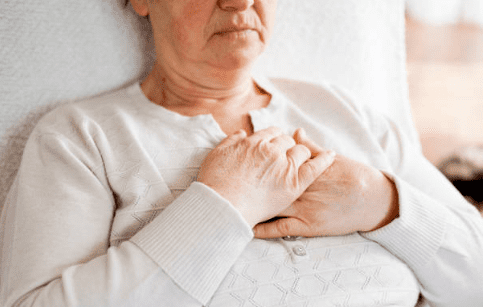
The testimonials and stories of heart transplant recipients give inspiration and hope to other patients waiting for this difficult process. Often these comments are filled with phrases such as “rebirth”, “the joy of being able to take a deep breath” or “the triumph of being able to climb stairs”. Patients return to a normal life where they can spend time with their loved ones, return to work, and even play sports, free from the limitations they experienced before the transplant, such as shortness of breath, constant fatigue, body aches and social withdrawal.
But this new life comes with certain rules. The immunosuppressive drugs used to prevent organ rejection (rejection) make the body more vulnerable to infections. Adopting a new lifestyle is therefore vital for the long-term success of the transplant.
Some key points to consider for a healthy life after heart transplantation:
- Timely and regular use of medicines
- Keeping doctor appointments and check-ups
- Paying utmost attention to personal hygiene
- Avoiding crowded and closed environments
- Avoiding contact with sick people
- Protect yourself from the harmful rays of the sun (use high-factor sunscreen)
Some important changes are also required in the diet. In general, the principles of healthy nutrition apply:
- Restrict salt consumption
- Avoiding processed and packaged foods
- Eat plenty of fresh fruit and vegetables
- Avoid raw or undercooked meat, eggs and unpasteurized dairy products
- Avoid grapefruit and pomegranate (they can interact with some medicines)
- Drink plenty of water
Physical activity is also an important part of life. After transplantation, light exercises started with the doctor’s approval are increased and continued over time. Activities such as regular walking, swimming or cycling are very good for both physical and mental health.

Prof. Dr. Yavuz Beşoğul graduated from Erciyes University Faculty of Medicine in 1989 and completed his specialization in Cardiovascular Surgery in 1996. Between 1997 and 2012, he served at Eskişehir Osmangazi University Faculty of Medicine as Assistant Professor, Associate Professor, and Professor, respectively. Prof. Dr. Beşoğul, one of the pioneers of minimally invasive cardiovascular surgery in Türkiye, has specialized in closed-heart surgeries, underarm heart valve surgery, beating-heart bypass, and peripheral vascular surgery. He worked at Florence Nightingale Kızıltoprak Hospital between 2012–2014, Medicana Çamlıca Hospital between 2014–2017, and İstinye University (Medical Park) Hospital between 2017–2023. With over 100 publications and one book chapter, Prof. Dr. Beşoğul has contributed significantly to the medical literature and is known for his minimally invasive approaches that prioritize patient safety and rapid recovery.