Doppler ultrasonography is a diagnostic imaging technique that measures blood flow in vessels using sound waves. It is widely used for vascular and cardiac evaluations.
The procedure is non-invasive and painless, involving a probe placed on the skin to visualize blood flow and detect narrowing, clots, or circulatory disorders.
Doppler ultrasound is essential in diagnosing deep vein thrombosis, carotid artery disease, and peripheral vascular disease. It is also used in pregnancy to assess fetal circulation.
Advantages of Doppler ultrasonography include its safety, absence of radiation, and real-time results. It is an effective, repeatable, and accessible diagnostic tool in modern medicine.
| Medical Name | Doppler Ultrasonography (Doppler USG) |
| Frequent Use Areas | – Vascular occlusions and stenoses – Vein and artery diseases (varicose veins, deep vein thrombosis, peripheral arterial disease) – Organ blood supply assessment |
| Causes | – Leg swelling, pain, suspicion of vascular disease – Evaluation of vascular structure and flow velocity |
| Risk Factors | – History of vascular diseases- Old age- Diabetes, hypertension, smoking |
| Complications | – No radiation, no serious complications – Rarely mild discomfort due to the procedure |
| Diagnostic Methods | – Clinical examination – Other imaging methods if necessary (CT, MR angiography) |
| Treatment Methods | – Doppler USG is for diagnosis and monitoring purposes, not a treatment method |
| Prevention Methods | – Regular vascular controls and follow-up in high-risk individuals |
What is Doppler Ultrasonography (Doppler USG) and How Is It Performed?
Doppler ultrasonography, or Doppler USG for short, is a painless and reliable imaging method that visualizes the movement, speed and direction of the blood flowing through our blood vessels using sound waves. You can think of this technology as the sound of an ambulance siren getting louder as it approaches us and fainter as it moves away. This basic principle of physics, the “Doppler Effect”, is used in medicine to measure the movement of blood cells. The probe of an ultrasound machine is both a source, sending out high-frequency sound waves, and a receiver, collecting the waves reflected from moving blood cells. The speed and direction of movement of the blood cells changes the frequency of the returned sound waves, and the device analyzes this change to give us invaluable information about vascular health. This procedure is performed completely externally, through the skin, without any incisions or radiation to the body.
What is the Difference Between Doppler and Normal Ultrasound?
Although these two terms are often used interchangeably, they are actually two separate technologies with different functions. In the diagnosis of vascular diseases, a combination of the two, i.e. “Duplex Ultrasonography”, gives us a complete picture and is therefore considered the gold standard.
Normal ultrasound, or B-Mode (Brightness Mode) ultrasound, produces the classic black-and-white image we all know. This method draws an anatomy map according to the density of the tissues. It shows the vessel itself, its wall structure, thickness and surrounding tissues as a two-dimensional photograph. But this photograph is static; it cannot tell what is happening inside it, whether blood is flowing or not, or how fast it is flowing.
Doppler ultrasound adds life and movement to this static map. Where normal ultrasound answers the question “Where is it?”, Doppler answers the question “What is happening?”. Doppler focuses on the movement of blood cells inside that vessel that we see with B-Mode. It measures the presence of blood, the direction in which it flows and, most importantly, how fast it moves. So one shows the structure of the vessel, the other shows its function, its physiology. One shows you a pipe, the other shows you the speed and flow pattern of the water through the pipe. The combination of the two allows us to see at the same time, for example, how much a vascular plaque (structural problem) is obstructing blood flow (functional problem).
What is looked for in a Doppler USG?
During a Doppler USG examination, many different parameters are focused on to assess the health of your vascular system from A to Z. This is a comprehensive analysis that involves much more than just looking at a single value. Basically, the information we are looking for is as follows:
- Presence or absence of blood flow
- Whether the blood flow is in the normal direction
- Speed of blood flow (slow, normal or extremely fast)
- Structure and thickness of the vessel wall
- Presence of plaques causing stenosis in the vessel
- Whether there is a clot in the vein
- Characteristic shape of the blood flow wave
- Flow changes before and after stenosis
What Are the Types of Doppler Ultrasonography and What Does It Do?
Just as a mechanic has different screwdrivers in his toolbox for different jobs, we have a variety of Doppler methods to examine different vascular problems. Each has its own advantages and limitations, and choosing the right method is the first step to a correct diagnosis.
Continuous Wave (CW) Doppler
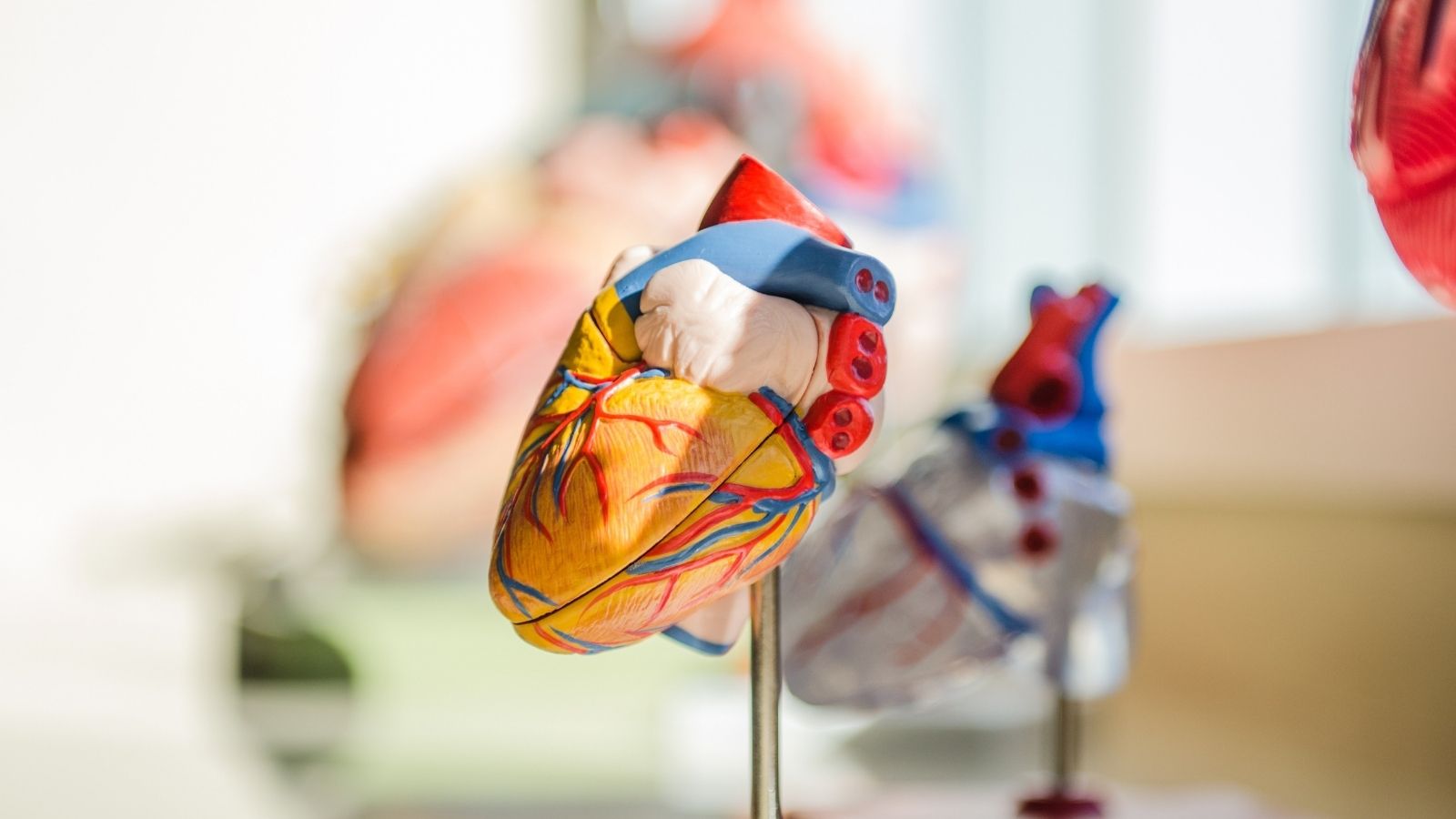
This is a simple but very powerful technique in which one crystal of the probe continuously sends out sound waves and the other continuously collects the reflections. It is used to accurately measure extremely high velocity blood flow, especially in severe stenosis or insufficiency of heart valves.
The advantages are as follows:
- It can accurately measure very high blood flow velocities.
- the speed measurement error called “aliasing” is not observed with this method.
But it has an important limitation:
It cannot determine the exact depth of the place where it is measuring the speed, i.e. it cannot distinguish which point on the road the signal is coming from.
Discrete Wave (PW) Doppler
This is the “workhorse” method we use most often in vascular examinations. A single crystal sends out short pulses of sound waves and then pauses to listen for returning echoes. This allows us to measure blood flow at a tiny point of our choosing, which we call the “sample volume”. This allows us to determine the exact location and degree of stenosis with incredible precision.
The most important advantage is this.
It allows the measurement of blood flow at a very specific point inside the vessel (range resolution).
But there is a limit:
When the blood flow rate exceeds a limit set by the device (Nyquist Limit), a measurement error called “aliasing” can occur, which makes it appear as if the current is flowing in the opposite direction.
Color Flow Doppler (CFD)
Color Doppler overlays blood flow as a colored map on a standard black-and-white ultrasound image. Traditionally, flow toward the ultrasound probe is coded in red and flow away from the probe is coded in blue. This is like a “blood flow road map” that allows us to understand at a glance where the vessels are, whether they are open or not, and the general pattern of blood flow. It allows us to immediately recognize problem areas such as stenosis or turbulence and guides us on where to place the PW Doppler for more detailed measurements.
The advantages are as follows:
- Instantly maps the direction and overall pattern of blood flow visually.
- It helps to quickly find problem areas such as stenosis or turbulence.
There are also some limitations:
- It does not provide a complete quantitative measurement as it only shows the average speed.
- By reducing the frames per second (frame rate) of the image, it can slightly reduce the sensitivity to time.
Power Doppler (PD)
Power Doppler works differently from other methods. It does not measure the speed or direction of blood flow, but its total strength – the amount of blood cells in motion. This is why it is usually expressed in a single color (often orange or yellow). Its greatest strength is that it can show even blood flows that are too slow or weak for other methods to detect.
The main advantages are:
- It is highly sensitive to very slow or low volume blood flow.
- The accuracy of the measurement is not as dependent on the angle of the probe to the vessel as other methods.
- the “aliasing” artifact does not occur in this mode.
The most important limitations are the following:
- It gives no information about the direction or speed of blood flow.
- Due to its high sensitivity, it is more prone to “flare” artifacts caused by the movement of surrounding tissues.
So, how is Doppler USG performed?
The answer to the question of how to perform a Doppler USG is to know that the procedure consists of systematic and painstaking steps. During the procedure, a water-based, transparent gel is applied to the skin in the area to be examined, which allows the sound waves to be transmitted to your body without airborne obstructions. The specialist then moves the ultrasound probe (transducer) over the skin to find the vessels. The vessels are examined in detail in both longitudinal and transverse sections. For example, in a leg artery examination, the entire vascular bed is followed step by step, starting from the main artery in the groin area to the thinnest vessels in the ankle. This systematic approach ensures that no vessel segment is missed and a comprehensive evaluation is performed.
Should I Be Hungry or Full for Doppler USG Before Examination?
This is one of the most common questions and the answer depends on which vascular area is to be examined. It is critical to follow some preparation rules to get accurate results.
In general, the preparation steps to be considered are as follows:
Abdominal Examinations: If intra-abdominal vessels such as the aorta, renal or mesenteric arteries are to be examined, a fast of 6 to 12 hours is requested before the procedure. This is to reduce gas in the intestines and to allow clear visualization of the deeper vessels. So the answer in this case is definitely to be “hungry”.
Nicotine and Caffeine Restriction: Smoking, nicotine patches, coffee, tea and cola should be stopped at least two hours before all Doppler examinations, especially when the arterial system is to be evaluated. These substances can cause constriction of blood vessels, artificially altering blood flow rates and leading to misinterpretation of the results.
Comfortable Clothing: No special preparation is usually required for areas such as the arms, legs or neck. You only need to wear loose and comfortable clothing that allows easy access to the area to be examined.
Pelvic Examinations: In some cases, especially to evaluate veins in the pelvis or early pregnancy ultrasounds, you may be asked to urinate and have a full bladder. The urine-filled bladder acts as an “acoustic window”, helping to provide a clearer view of the structures behind it.
Specifically, How to Take a Leg Doppler?
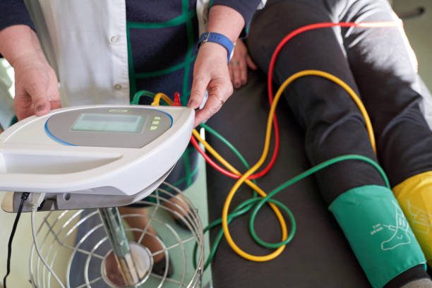
to better understand the question “How is a leg Doppler performed?”, we can consider one of the most basic screening tests for atherosclerosis, the Ankle-Arm Pressure Index (ABPI). Even this simple test shows how important a standardized protocol is.
The patient is first placed on their back in a warm room and rested for about 10-15 minutes. This rest period allows the body to return to its basal state, removing the temporary effects of a nearby walk or excitement on blood pressure. Blood pressure is then first measured in both arms. To do this, a sphygmomanometer cuff is wrapped around the arm and a Doppler probe is placed on the artery at the elbow. As the cuff is inflated and deflated, the pressure reading at which the first sound is heard when blood flow resumes is recorded. This process is repeated on both arms and the highest pressure value is taken as reference.
The same procedure is then repeated for the legs. This time the sphygmomanometer cuff is wrapped just above the ankle. With the Doppler probe, pressures are measured separately in the arteries in the dorsum of the foot and in the inner part of the ankle. Finally, for each leg, the highest pressure measured at the ankle is divided by the highest pressure measured at the arm. A ratio between 1.0 and 1.4 is considered normal, while values below 0.9 indicate stenosis or occlusion of the leg arteries (peripheral arterial disease).
How Many Minutes Does a Doppler Ultrasound Take?
There is no clear answer to this question, because the answer to the question of how many minutes Doppler ultrasound takes depends entirely on the purpose and scope of the examination. For example, a venous Doppler examination to investigate a suspected clot (DVT) in a single leg can usually be completed in 15-30 minutes. However, complex procedures such as a comprehensive arterial Doppler, where both leg arteries are assessed in detail from groin to ankle, or vascular mapping for a pre-operative bypass can take 60 to 90 minutes or longer. The duration may also depend on how easily the patient’s vasculature can be visualized and the complexity of the findings.
What do the waveforms in the Doppler result mean?
One of the most valuable outputs of Doppler examination is a graph called a “spectral waveform”. This graph is like an ECG of the blood flow, showing not only its speed but also its character. The shape of the wave gives us valuable information about whether there are any obstacles in the path of the flow and the condition of the tissue the blood is traveling through.
Here are some typical waveforms we see in healthy and problematic vessels:
Triphasic wave: This is the healthy waveform of a normal arm or leg artery at rest. It consists of a sharp forward current with the heartbeat, a short backward current caused by the elastic recoil of the vessel wall and finally a small forward current phase. This indicates that the vessels at the ends have a healthy resistance to blood flow.
Biphasic wave: Forward and reverse current phases are present, but the third phase is lost. Sometimes it can be a normal variation, sometimes it is an early sign of vascular disease.
Low Resistance Monophasic Wave: This waveform shows a sharp peak followed by a continuous forward current throughout the cardiac cycle. This is normally normal in vessels supplying organs that need constant blood supply, such as the kidneys or brain. However, it is abnormal if it occurs in the arteries of a leg or arm and usually indicates a condition that causes the veins to dilate, such as an infection, trauma or exercise in that area.
Damped Monophasic (Tardus Parvus) Wave: This is one of the most important pathologic findings for us. The waveform is weak, rounded and delayed in reaching its peak. This image is a sure sign that there is a significant stenosis or complete blockage behind the point of measurement, i.e. in the direction of the blood flow, which severely obstructs the flow.
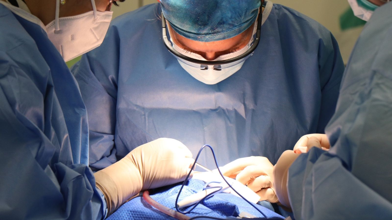
Why is Doppler Ultrasonography so Important in Surgery?
Doppler USG is a cardiovascular surgeon’s closest ally. It transforms surgery from an unknown into a precise and targeted intervention. It plays an indispensable role in both preoperative planning and postoperative follow-up.
The main purposes of use in the preoperative period are as follows:
Vein Mapping for Bypass: Determines the location, diameter, quality and health of the best vein to use for bypass surgery (usually the patient’s own leg or arm vein) before surgery. This avoids unnecessary large incisions, shortens the operation time and reduces the risk of complications.
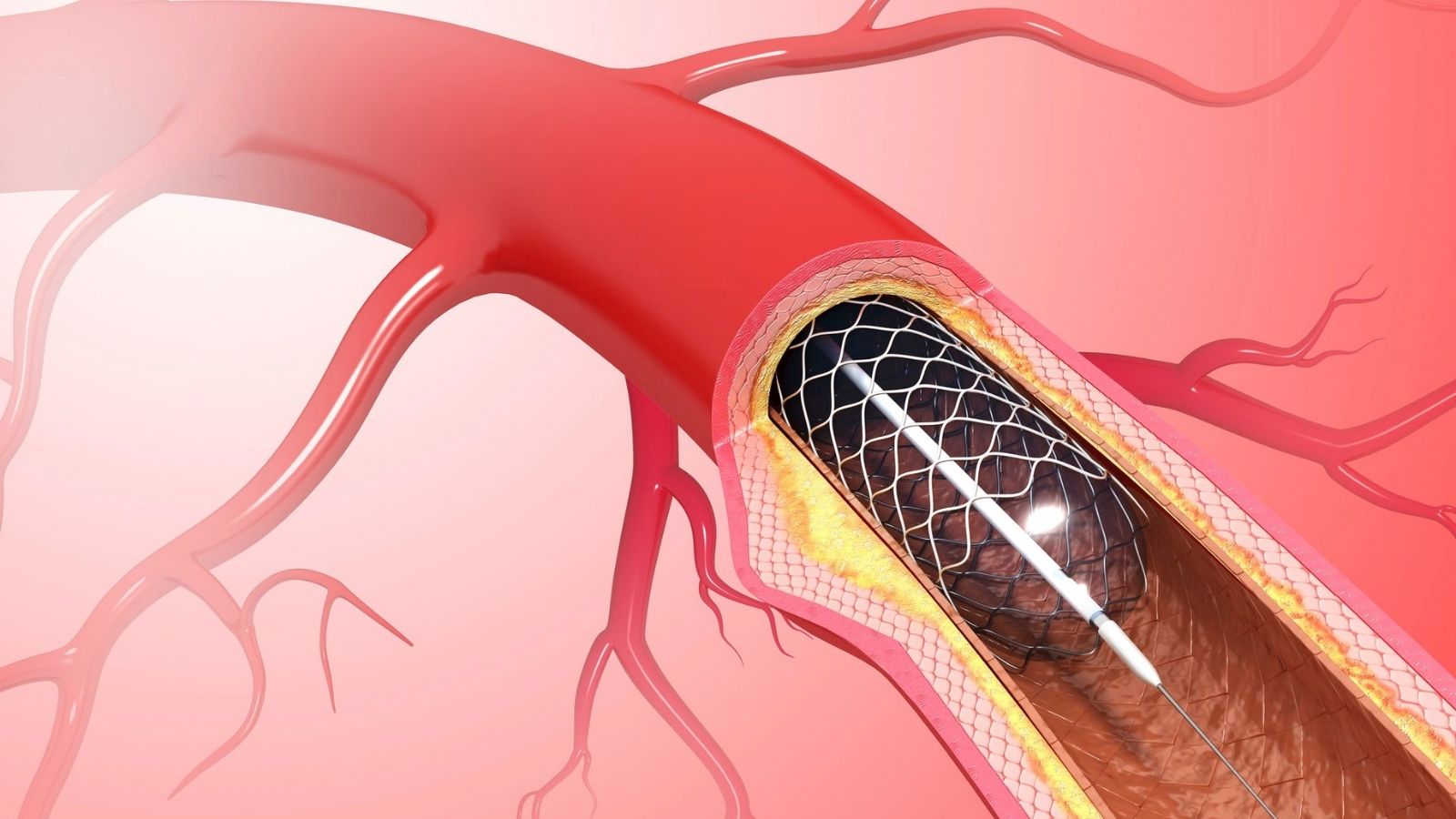
Determine the exact location and severity of the stenosis: It clearly shows which vessel has a stenosis and how severe it is, allowing us to choose the right treatment method (balloon, stent, plaque removal or bypass).
Evaluation of the Inflow and Outflow Vessels: For a bypass graft to be successful, the “inflow” vessel to bring blood in and the “outflow” vessel to take blood out must be healthy. Doppler checks the compliance of these target vessels.
Examination of the Structure of Vascular Plaques: It allows us to plan our maneuvers during surgery more safely by showing whether the plaque causing the stenosis is “soft” and can easily clot or “hard” and calcareous.
Its role in postoperative follow-up is just as vital.
Control of Grafts and Stents: It can detect the re-narrowing (restenosis) or blockage of bypass grafts or stents at a very early stage, often without the patient feeling any discomfort.
Follow-up After Aneurysm Repair: After closed repair of an aortic aneurysm, the stent checks for blood leaks (endoleaks) around the graft. These leaks mean that the aneurysm is still at risk of bursting.
Verification of Treatment Success: It objectively shows whether the blood flow improves after the intervention and whether the velocities return to normal.
Monitoring the Diameter of the Aneurysm Sac: After a successful aneurysm repair, the aneurysm sac is expected to shrink over time. The Doppler confirms the success of the treatment by following this shrinkage with regular measurements.
Which Vascular Diseases Can Doppler Diagnose?
Doppler ultrasonography is used as the primary method for diagnosing a wide range of vascular diseases.
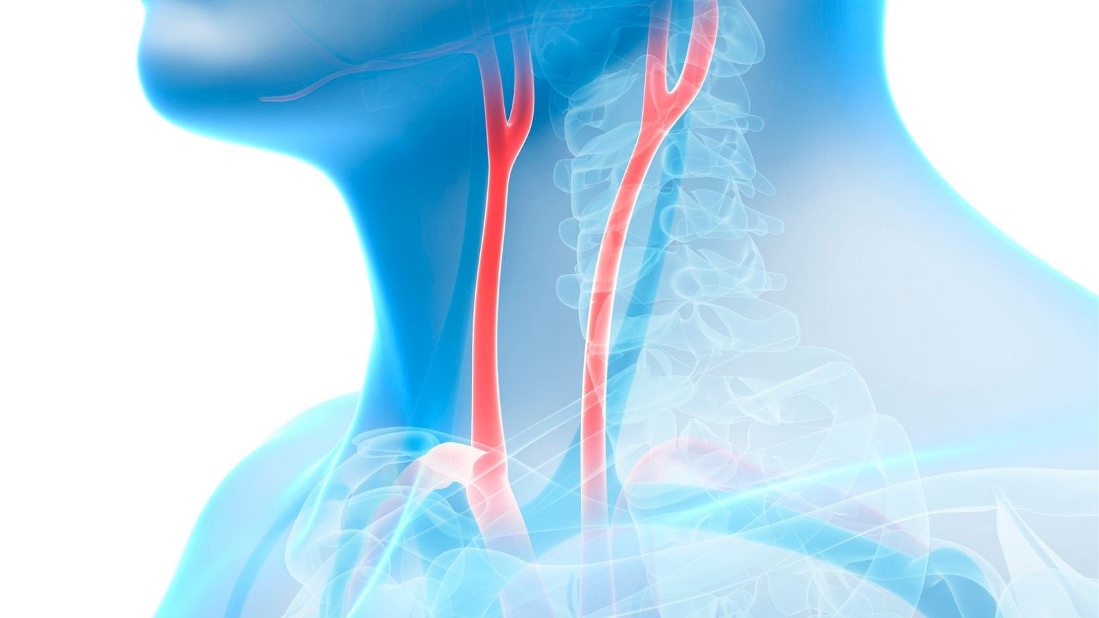
Carotid Artery Stenosis (Neck Carotid Stenosis)
It is the gold standard for detecting and grading stenosis in these critical vessels that carry blood to the brain. It plays a vital role in determining the risk of stroke.
The main findings are as follows:
- Calcification or plaque structures in the vessel wall visible with B-mode.
- Significant increase in blood flow velocity at the site of stenosis (PSV > 230 cm/s for severe stenosis).
- Marked “spectral broadening” of the flow wave indicating turbulence.
- In the case of a complete blockage, no blood flow signal can be received within the vessel.
Peripheral Arterial Disease (Arterial Stenosis of the Arteries of the Arm and Leg)
It is used to determine the location and severity of stenosis and blockages, especially in the leg veins. It identifies the cause of complaints such as walking pain (claudication) or pain at rest.
The main findings are as follows:
- Loss of the healthy, three-phase (triphasic) flow wave that should normally be present in the leg.
- Presence of a “damped” (Tardus Parvus) monophasic waveform in vessels beyond the stenosis.
- More than 2-fold increase in velocity at the point of stenosis compared to the immediately preceding normal zone (PSV Ratio > 2.0).
- In complete occlusions, there is no flow in that area and weak “collateral” (side branch) flows are observed immediately after the occlusion.
Deep Vein Thrombosis (DVT – Clot Formation in Vein)
It is the most reliable method for diagnosing this dangerous condition, which occurs especially in the deep veins in the legs and carries the risk of clotting in the lung (pulmonary embolism).
The main findings are as follows:
The most important finding: When the vessel is gently pressed with the ultrasound probe, the walls of the clot-filled vessel do not fuse, i.e. do not “collapse”, unlike in a healthy vessel.
- Direct visualization of the clot in the vessel lumen.
- No flow signal in that area on color Doppler.
- No acceleration of blood flow in the vein above (no augmentation) when the calf is gently pressed.
Chronic Venous Insufficiency (Internal Varicose Veins or Vein Valve Insufficiency)
It is used to diagnose deterioration of the vein valves and backward leakage of blood (reflux), which causes swelling, pain, discoloration and sores in the legs.
The main findings are as follows:
- When the patient is standing or when special maneuvers (such as squeezing and releasing the calf) are performed, the blood escapes backwards, i.e. towards the foot, contrary to what should normally happen.
- This retraction time is pathologically long (usually > 0.5 seconds).
- Enlargement of the diameter of the insufficient vessels.
- Direct visualization of impaired valve structures.

Prof. Dr. Yavuz Beşoğul graduated from Erciyes University Faculty of Medicine in 1989 and completed his specialization in Cardiovascular Surgery in 1996. Between 1997 and 2012, he served at Eskişehir Osmangazi University Faculty of Medicine as Assistant Professor, Associate Professor, and Professor, respectively. Prof. Dr. Beşoğul, one of the pioneers of minimally invasive cardiovascular surgery in Türkiye, has specialized in closed-heart surgeries, underarm heart valve surgery, beating-heart bypass, and peripheral vascular surgery. He worked at Florence Nightingale Kızıltoprak Hospital between 2012–2014, Medicana Çamlıca Hospital between 2014–2017, and İstinye University (Medical Park) Hospital between 2017–2023. With over 100 publications and one book chapter, Prof. Dr. Beşoğul has contributed significantly to the medical literature and is known for his minimally invasive approaches that prioritize patient safety and rapid recovery.