Atrial septal defect (ASD) is a congenital heart anomaly characterized by an opening in the wall between the atria. This defect allows oxygenated and deoxygenated blood to mix, potentially causing enlargement of the right heart chambers over time.
Small defects may remain asymptomatic, while larger ones can lead to fatigue, shortness of breath, and recurrent respiratory infections. If untreated, they may progress to pulmonary hypertension or heart failure in adulthood.
Diagnosis is usually made through echocardiography, which visualizes the septal defect and measures its size. Additional imaging or cardiac catheterization may be required for detailed assessment, especially before surgical planning.
Treatment depends on the size and impact of the defect. While small ASDs may close spontaneously, larger defects often require closure via catheter-based interventions or open-heart surgery to prevent long-term complications.
| Other Names | Atrial Septum Defect, Hole in the heart (between the atria) |
| Affected System | Cardiovascular System (heart) |
| Causes | Congenital; septal closure defect in the fetal heart |
| Types | Secundum ASD (most common), Primum ASD, Sinus venosus type, Coronary sinus type |
| Symptoms | Usually asymptomatic in childhood; rapid fatigue, shortness of breath, palpitations, frequent respiratory tract infections in adults |
| Diagnostic Methods | ECG, echocardiography (transthoracic or transesophageal), cardiac catheterization, MRI |
| Complications | Right heart failure, arrhythmias, stroke risk (paradoxical embolism), pulmonary hypertension |
| Treatment Methods | Small defects may be observed in infancy; immediate surgery when detected at older ages. Surgical repair is performed with a minimal incision in the armpit. |
| Age at Intervention | It can be performed in symptomatic childhood, usually between 2-5 years of age in asymptomatic cases, and in adulthood in late cases |
| Follow-up and Monitoring | Echocardiography control in the first year postoperatively. |
| Prognosis | He is very well after the operation, he continues his normal life. |
What is Atrial Septal Defect and How Does It Affect the Heart?
To fully grasp this, let’s take a look at the magnificent architecture of our heart. You can think of the heart as a two-storey house with two separate apartments next to each other. On the upper floors of this house are the “atria” (atria) and on the lower floors are the “ventricles” (ventricles). The chamber on the right side of the house circulates around the body, collects tired, deoxygenated blood and sends it to its next-door neighbor, the lungs, to be cleaned. Freshened with oxygen in the lungs, the blood returns to the apartment on the left side of the house, where it is pumped vigorously and distributed throughout the body.
Between these two circles, between the right and left sides of the heart, there is a thick wall that prevents the mixing of clean and dirty blood. In medicine, we call this wall the septum. The answer to the question of what is the heart septum is the wall of the heart that separates this life. Atrial Septal Defect is a hole in the wall (atrial septum) between the atria, which are the upstairs rooms of this house. So when asked what a s d means, the answer is the hole in the wall between the atria.
What causes this hole? The pressure on the left side of the heart is always higher than on the right side. Because of this pressure difference, some of the oxygen-rich clean blood that should normally go to the whole body escapes through this hole to the right side. This abnormal passage of blood causes dirty blood to mix with clean blood coming into the right atrium, and the total amount of blood sent to the lungs becomes much higher than normal.
This is similar to a water pump designed for watering a garden, trying to water the garden and fill the pool at the same time. The pump (the right side of the heart) has to work much harder than normal. Over the years, this extra load tires and expands the pump’s motor and degrades its performance over time. This is exactly what ASD does to the heart. The right side of the heart gradually becomes larger and fatigued, and in the long term leads to increased pressure in the pulmonary arteries (pulmonary hypertension) and heart failure.
How Many Different Types of Atrial Septal Defects Can Be Seen?
The Atrial Septal Defect cases we encounter differ according to the location of the hole on the atrial septum. This classification plays a critical role in determining our treatment approach. The main types of ASD are:
- Secundum ASD
- Primum ASD
- Sinus Venosus ASD
- Coronary Sinus Defect
Let’s take a closer look at what these types mean. Secundum ASD is the most common type, accounting for more than three quarters of all cases. The location of the hole is right in the middle of the septum. The most important feature of this type is that it is usually not associated with another heart abnormality and, in structurally appropriate cases, it is highly amenable to catheter closure, popularly known as the “umbrella method”. This is a great comfort and advantage for patients.
Primum ASD is located in the lower part of the septum, very close to the valves between the upper and lower chambers of the heart. Because of this location, it is often found in combination with additional valve problems, such as a mitral valve defect. Therefore, its treatment always requires surgical repair, during which both the hole is closed and the problem with the valve is corrected.
Sinus Venosus ASD is a rarer type. It is located at the upper or lower end of the septum, near the entrance to the large veins (vena cava) that bring the main blood supply to the heart. An important feature of this type of ASD is that one or more of the veins carrying clean blood from the lungs often mistakenly open into the right atrium instead of the left (abnormal pulmonary venous return). This additional condition necessitates surgical repair and these vessels need to be directed to the correct location during surgery.
Finally, the rarest type, Coronary Sinus Defect, is usually seen as part of much more complex congenital heart diseases and its treatment is planned according to this complex condition.
There is also Patent Foramen Ovale (PFO), which is often confused with ASD. PFO is when a natural valve, which is present in every baby in the womb and should close at birth, remains open. PFO is not a true tissue defect, a hole, like ASD. This condition, which occurs in about a quarter of the population, is usually innocent and does not require treatment.
Why Does Atrial Septal Defect Occur and Who is at Risk?
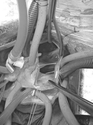
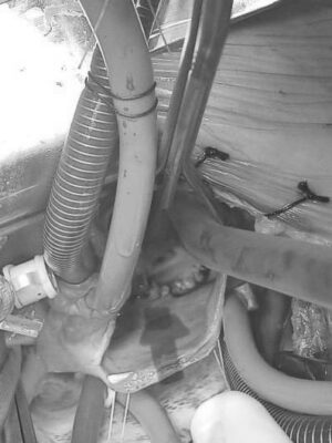
Atrial septal defect, a hole in the atrial septum between the atria of the heart[/caption
First of all, the most important point must be emphasized: Atrial Septal Defect is not caused by a mistake made by the mother or father. It is a defect that occurs during the development of the baby in the womb, at a very delicate stage in the formation of the heart. Although the exact causes are often unknown, it is thought that certain factors may increase this risk.
Genetic factors can play an important role. A family history of congenital heart disease may slightly increase the risk. In addition, babies with certain genetic syndromes such as Down syndrome and Holt-Oram syndrome are more likely to have ASD.
Some conditions that the mother encounters during pregnancy can also affect the development of the baby’s heart, increasing the risk slightly. Some of these factors:
- Rubella infection
- Uncontrolled diabetes
- Autoimmune diseases such as lupus
- Some antiepileptic drugs
- Lithium-containing drugs
- Alcohol consumption
- Smoking
- Drug use
What are the Most Common Atrial Septal Defect Symptoms?
Symptoms of ASD vary greatly depending on the size and type of hole and, most importantly, the age of the person. A small hole can remain unnoticed for a lifetime, while a large hole can cause significant symptoms over time. For this reason, it is more accurate to analyze the symptoms under two separate headings: childhood and adulthood.
During infancy and childhood, especially if the hole is large, here are some of the symptoms that an Atrial Septal Defect can show:
- Quick fatigue
- Shortness of breath (especially during activity)
- Inadequate weight gain
- Slow growth
- Recurrent lung infections (bronchitis, pneumonia)
- Feeding difficulties (in infants)
- Audible heart murmur
The most common finding in children is a “murmur” heard with a stethoscope during a routine examination. This abnormal sound is often the first sign that leads a family to a pediatric cardiologist. Other symptoms indicate that the heart and lungs are struggling to cope with the increased blood flow. Growth retardation and failure to gain weight can occur because the energy the body needs for growth and development goes to the heart, which is constantly working overtime. The constant “wetness” of the lungs makes them more prone to infections.
Atrial Septal Defect, which can progress without symptoms for years, usually manifests itself in adulthood with the following complaints:
- Shortness of breath with exertion
- Chronic fatigue
- Palpitations
- Irregular heartbeat
- Swelling in the feet, legs or abdomen (edema)
- Susceptibility to stroke
- Migraine headaches
- Bruising of the lips (in advanced cases)
These symptoms in adulthood are a sign that the heart and lungs can no longer withstand the strain they have been under for years. The most common complaints are breathlessness, even when climbing stairs or doing simple housework, and a constant state of exhaustion throughout the day. Palpitations and arrhythmias, especially after the 40s, are the result of the atria of the heart stretching and enlarging over the years. Swelling in the feet is an important warning sign of the onset of right heart failure.
Which Methods Are Used to Diagnose Atrial Septal Defect?
When ASD is suspected, we perform a series of evaluations to confirm the diagnosis and plan treatment properly. This process is similar to detective work, with each test completing an important piece of the puzzle.
The diagnostic journey usually starts with physical examination. When we listen to the heart with a stethoscope, findings such as “fixed paired second heart sound” and “systolic murmur”, which are very typical for ASD, are the first clues that point us in the right direction.
However, the most valuable method that confirms the diagnosis and draws our roadmap is Echocardiography (ECHO), an ultrasound of the heart. This painless and radiation-free test provides a detailed map of the heart. With a standard ECHO (Transthoracic ECHO) through the chest wall, we can clearly see the presence, location and size of the hole, how dilated the chambers of the heart are and how much blood is passing through the hole. Sometimes, especially in adults or when catheter closure is planned, Transesophageal Echocardiography (TEE) through the esophagus may be needed to get clearer images. This method gives us invaluable information to assess the edge structure of the hole and whether the closure device can be placed safely.
Although echocardiography largely confirms the diagnosis, additional tests may be needed to fully assess the condition:
- Electrocardiogram (ECG)
- Chest X-ray (Chest X-ray)
- Cardiac Magnetic Resonance (MRI)
- Computed Tomography (CT)
- Cardiac Catheterization
An ECG records the electrical activity of the heart, showing rhythm disturbances or overload on the right side of the heart. A chest X-ray gives an idea of the overall size of the heart and increased blood supply to the pulmonary vessels. Cardiac MRI or CT is used to measure the volume and function of the heart chambers very precisely, especially when the anatomical structure is complex or echocardiography is inadequate.
Cardiac Catheterization is an interventional procedure. We use it before deciding on treatment, especially in older patients or in patients with suspected pulmonary hypertension. In this procedure, thin tubes are inserted into the heart through a vein in the groin and the intracardiac pressures and oxygen levels in the blood are measured directly. This gives us the most precise information about whether closing the hole would be beneficial or harmful for the patient.
What Treatment Options Are Available for Atrial Septal Defect?
When an atrial septal defect is diagnosed, regardless of age, the treatment is surgical. It is rarely closed with a very small asd catheter and metal plates called the umbrella method.
Observation and Medical Monitoring
It is known that some of the very small holes (less than 4 mm) detected in infancy close spontaneously within the first few years. In this case, it is sufficient to invite the patient for cardiology check-ups at regular intervals and monitor the situation closely.
Complications such as displacement of the device, abrasion of the heart wall, clot formation or rhythm problems can occur with the Umbrella with Catheter method. Therefore, it is vital to choose the right patient and have the procedure performed by an experienced team. Blood thinners (such as aspirin) are usually used for 6 months after the heart umbrella is implanted, until the device is covered with tissue.
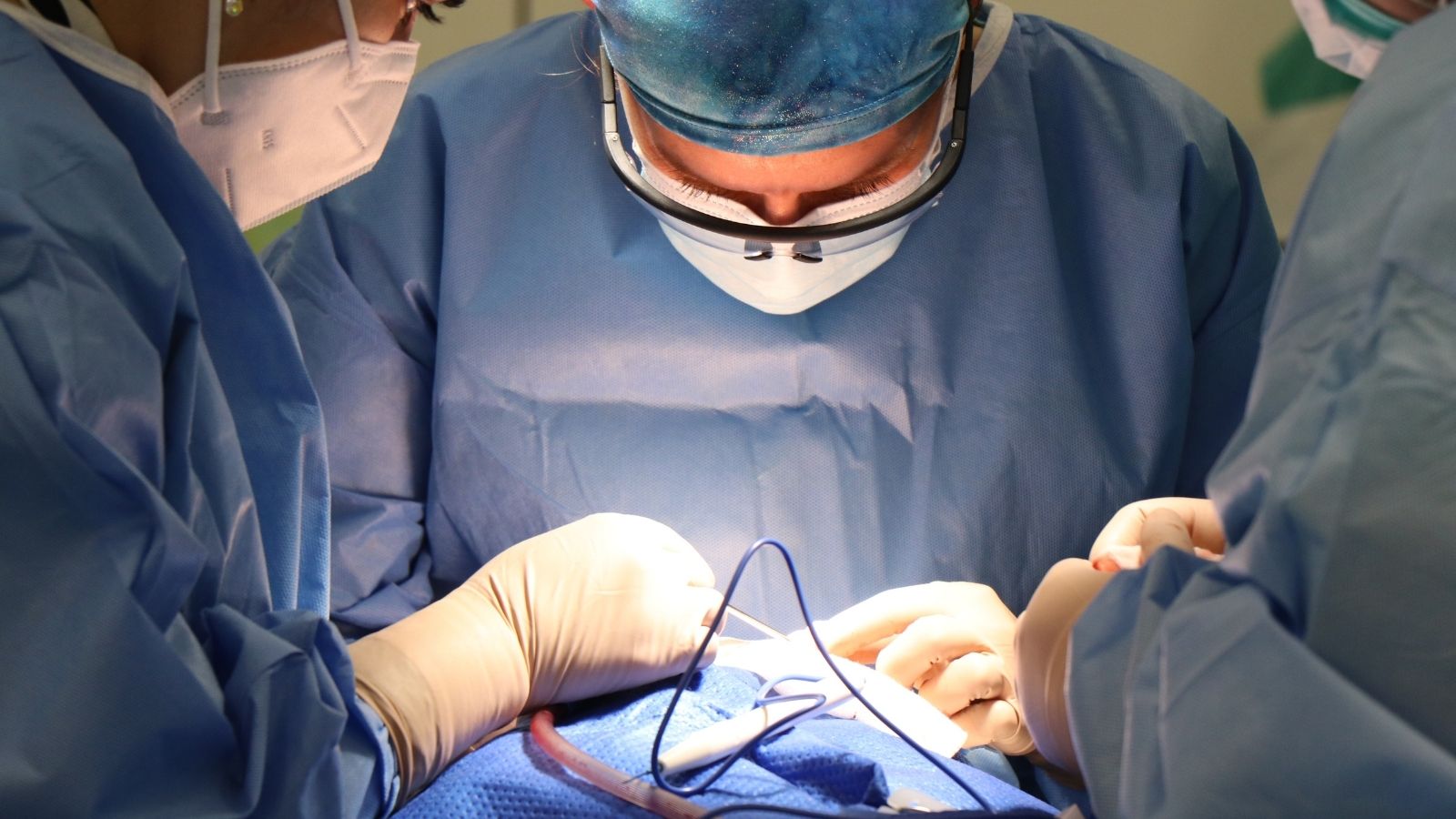
Surgical Treatment
Surgical repair remains the gold standard and definitive solution for patients who are not amenable to catheterization or who have additional cardiac problems. ASD types such as primum, sinus venosus or secundum ASDs that are too large to be closed by catheter or have insufficient margin tissue are treated surgically.
Today, minimally invasive surgery is performed in the armpit to access the heart and, with the support of a heart-lung machine, the hole is closed either directly with sutures or with a synthetic patch or a patch made from the patient’s own heart membrane.
Risks “Is there a risk of death with heart hole surgery?” is a major concern for families. In experienced centers, the risk of surgery to repair an isolated ASD in a patient with no other serious health problems is extremely low (less than 0.5%). This risk may be even lower than the risk of appendectomy.
The timing of treatment is also important. Generally, it is preferable to close the hole before school age (between 3-5 years), before irreversible changes in the heart and lungs begin. However, when diagnosed in adulthood, surgery is recommended immediately. What Problems Can an Untreated Atrial Septal Defect Cause?
While small and insignificant holes are usually not a problem, an untreated medium to large Atrial Septal Defect can lead to serious and life-threatening complications in the long term. Therefore, closing a significant hole, even if there are no symptoms, is a proactive health investment. The main problems that can be encountered when left untreated are:
- Right Heart Failure
- Pulmonary Hypertension (Lung Hypertension)
- Eisenmenger Syndrome
- Heart Rhythm Disorders (Arrhythmias)
- Paradoxical Embolism (Stroke Risk)
- Decreased Life Expectancy
Right heart failure occurs when the right ventricle, which is constantly overworked, gradually tires and loses its contractile strength, leading to fluid accumulation in the body (edema), shortness of breath and severe weakness. Pulmonary hypertension is the hardening and narrowing of the pulmonary vessels as a result of the constant pumping of blood at high pressure into the lungs. When this condition progresses, the pressure in the right heart exceeds that in the left heart and the direction of blood flow reverses. This is called Eisenmenger’s Syndrome and it is no longer possible to close the hole after this stage.
Rhythm disturbances, especially atrial fibrillation, are very common in patients over the age of 40 with untreated ASD. This condition, caused by enlarged and malformed atria, leads to palpitations and, more importantly, to the formation of clots inside the heart. Paradoxical embolism is when small clots that form in places such as the veins of the legs pass through the ASD hole and travel to the brain, causing a stroke. All these complications can negatively affect life expectancy in individuals with untreated significant ASD.
After ASD Surgery
| Recovery Time | usually 10-15 days after closed minimally invasive surgery |
| Type of Surgery | repair with closed heart surgery or closure with a catheterized device (percutaneous method). |
| Physical Activity | limited activity in the first weeks; return to normal activity is possible in a short time after catheterization. |
| Medication Use | Anticoagulants (especially if a device is implanted), aspirin, rhythm regulators can be used on doctor’s recommendation. |
| Rhythm Monitoring | Rhythm disorders (especially atrial fibrillation) can be monitored; ECG and Holter monitoring are performed. |
| Imaging and Control | Cardiac function is monitored after closure by regular echocardiography; more frequent in the first year. |
| Complications | Rhythm disturbances, device displacement (umbrella method), pericarditis, rarely residual shunt. |
| Wound and Catheter Site Care | After minimally invasive surgery or inguinal catheter site should be kept clean and dry. |
| Nutrition | No special diet may be required, but a heart-healthy diet is recommended. |
| Smoking and Alcohol | Smoking is strongly discouraged; alcohol consumption should be limited. |
| Sexual Activity | It can usually be started within 2-4 weeks once physical competence is achieved. |
| Psychological Support | Support may be needed for psychological adaptation, especially in individuals who had surgery at a young age. |
| Vehicle Use | It can usually be started 2-4 weeks after open surgery and 1 week after percutaneous intervention. |
| Controls | Regular cardiology check-ups are recommended during the first year and annually thereafter. |
Frequently Asked Questions
What happens if ASD is not closed?
Over the years, an ASD (hole between the upper chambers of the heart) tires and enlarges the right side of the heart and the pulmonary artery. In later life, it can lead to serious problems such as arrhythmias, heart failure and high pulmonary blood pressure. Early intervention is the best way to protect your heart.
By what age is ASD closed?
Most small ASDs can close spontaneously within the first 2 years of life. This is why we usually prefer to monitor them. After the age of two, large ASDs are very unlikely to close spontaneously and often require intervention.
Does ASD prevent military service?
It depends on the size of the ASD and its effect on the heart. Small ASDs that do not affect the heart may not prevent military service. However, if the heart is enlarged or has significant problems, the ASD is usually considered a disqualification for military service. For ASDs that are successfully closed, a detailed evaluation is performed again.
Does ASD cause pain?
ASD does not directly cause pain. The most common complaints are fatigue, shortness of breath or palpitations. If you have chest pain, other causes should be investigated.
What is an ASD murmur?
The murmur heard in ASD is a soft, characteristic sound made by ascending blood as it passes through the pulmonary artery. This sound is the typical first clue of ASD and is confirmed by echocardiography (ECHO).
How many mm should the ASD be?
The ideal is zero mm, but what really matters is the load the hole places on the heart and whether the chambers are enlarged. In general, ASDs that exceed 8-10 mm or cause heart enlargement require treatment.
ASD or VSD more dangerous?
The danger of each varies according to size and location. VSDs can cause problems earlier in infancy, while ASDs can progress insidiously over the years and cause problems later in life. Both require specialist follow-up.
Is ASD genetic?
Most ASDs occur by chance, but sometimes they run in families. If you have a first-degree relative with ASD, your risk may increase slightly, but there is no rule that it will be certain.
How is ASD diagnosed?
First, a typical murmur is heard on examination. The definitive diagnosis is made by echocardiography (ECHO). ECHO is a painless ultrasound test that shows the location and size of the hole and its effect on the heart.
When does ASD spontaneously shut down?
Small ASDs, especially in the middle wall (secundum ASD), usually close within the first 1-2 years of life. Large or different types of ASDs usually do not close spontaneously and require treatment.

Prof. Dr. Yavuz Beşoğul graduated from Erciyes University Faculty of Medicine in 1989 and completed his specialization in Cardiovascular Surgery in 1996. Between 1997 and 2012, he served at Eskişehir Osmangazi University Faculty of Medicine as Assistant Professor, Associate Professor, and Professor, respectively. Prof. Dr. Beşoğul, one of the pioneers of minimally invasive cardiovascular surgery in Türkiye, has specialized in closed-heart surgeries, underarm heart valve surgery, beating-heart bypass, and peripheral vascular surgery. He worked at Florence Nightingale Kızıltoprak Hospital between 2012–2014, Medicana Çamlıca Hospital between 2014–2017, and İstinye University (Medical Park) Hospital between 2017–2023. With over 100 publications and one book chapter, Prof. Dr. Beşoğul has contributed significantly to the medical literature and is known for his minimally invasive approaches that prioritize patient safety and rapid recovery.