The iliac artery is a major blood vessel that carries oxygenated blood from the abdominal aorta to the lower limbs. It divides into internal and external branches, each serving specific regions, and plays a vital role in ensuring proper circulation to the pelvic organs and legs.
Iliac artery anatomy is characterized by its bifurcation from the abdominal aorta, usually at the level of the fourth lumbar vertebra. The right and left iliac arteries follow symmetrical paths, allowing balanced blood distribution and maintaining vascular stability in the pelvic region.
The function of the iliac artery primarily involves supplying oxygen-rich blood to lower extremities. The internal iliac branch nourishes pelvic organs, bladder, and reproductive structures, while the external iliac continues as the femoral artery, ensuring perfusion of the leg muscles and skin.
Iliac artery pathologies include stenosis, aneurysm, and thrombosis. These conditions may cause leg pain, impaired circulation, and tissue ischemia. Early diagnosis through imaging and prompt vascular intervention are essential to prevent severe complications such as limb loss.
| Medical Name | Iliac Artery |
| Anatomical Location | – Originates from the lower part of the abdominal aorta, divided into right and left – Carries blood to the pelvis and legs |
| Duties | – Provides oxygenated blood to the pelvis and lower extremities (legs) |
| Common Diseases | – Iliac artery occlusion or stenosis – Aneurysm (ballooning) – Atherosclerosis (arteriosclerosis) |
| Risk Factors | – Hypertension- High cholesterol- Diabetes- Smoking- Advanced age |
| Complications | – Circulatory disturbance in the lower extremities – Walking pain in the legs (claudication) – Tissue loss and ulcer development |
| Diagnostic Methods | – Doppler ultrasonography- CT angiography- MR angiography- Angiography |
| Treatment Methods | – Drug treatment (blood thinners, statins)- Angioplasty and stenting- Bypass surgery (in advanced cases) |
| Prevention Methods | – Healthy diet – Not smoking – Regular exercise – Blood pressure and cholesterol control |
What Is the Iliac Artery and Why Is It So Vital for Our Body?
The iliac artery is the name given to the beginning of the arteries of the right and left leg, formed by the bifurcation of the last part of the main artery of the aorta. Think of it this way; the trunk of a large tree (the aorta) first splits into several main branches to deliver the water it receives from the soil to the leaves at the top. The iliac arteries serve as these main branches for the lower half of our body. Thanks to these branches, blood is both distributed to the vital organs in our pelvis (pelvis) and delivered uninterruptedly to the very ends of our legs. The proper functioning of this system underpins everything in our daily lives, from the simplest movements to the most complex functions.
Here are some of the basic functions we perform thanks to the blood flow provided by the iliac arteries:
- Walking and running
- Climbing stairs or hills
- Bladder (urinary bladder) functions
- Bowel movements and defecation
- Sexual function
- Nutrition of the hip and leg muscles
- Health of the skin and bones of the legs
What is the common iliac artery and what is its role in circulation?
The first and largest vessel section immediately after the bifurcation of the aorta is called the common iliac artery. There are two, one on the right and one on the left. You can think of these vessels as the first main exits of a large highway. Their job is to safely transfer the high-pressure blood they receive from the aorta to the smaller roads that branch off a little further down the highway – the internal and external iliac arteries. These vessels are located quite deep in the body, close to the back wall of the abdomen. This deep location and the important structures around them make any intervention in this area very delicate. For a surgeon, working in this region is like working at an intersection with heavy traffic and critical infrastructure lines.
There are some critical structures in close proximity to the main iliac artery:
- Iliac veins (veins)
- Ureter (urinary ducts from the kidney)
- Regional lymph nodes
- Autonomic neural networks
- Psoas muscle (the large muscle that connects the waist and buttocks)
What is the Role of the Internal Iliac Artery in Pelvic Nutrition?
After traveling a short distance, the main iliac artery splits again. One of these new pathways leads into the pelvic cavity (pelvis). We call this vessel the internal iliac artery. This vessel is the lifeblood of the pelvic region. It is responsible for the nutrition of all organs and structures in the pelvis. Without this vessel, our reproductive and excretory systems would not function. Especially in women, the “uterine artery”, the branch that feeds the uterus, miraculously expands and increases blood flow to meet the needs of the baby during pregnancy. We can see from this how vital this vessel is.
The main organs and sites to which the internal iliac artery carries blood are
- Bladder (urinary bladder)
- The lower part of the rectum (the last part of the large intestine)
- Prostate (in men)
- Uterus (in women)
- Vagina (in women)
- Hip joint and surrounding muscles
- Perineum and external genital organs
What is the Importance of the External Iliac Artery in Blood Flow to the Legs?
The other, larger branch of the main iliac artery is called the external iliac artery. Instead of entering the pelvis, this artery continues on its way to the groin area. At the point where it passes under the inguinal ligament and enters the leg, its name changes and it is now known as the femoral artery. The femoral artery is the main artery of the entire leg. So the external iliac artery is the starting point, the first and most important part of this super highway that carries blood to our legs. When we walk, when we play sports, all the oxygen and energy that our muscles need is transported through this artery. Even the slightest narrowing of this vessel directly affects our walking capacity and severely reduces our quality of life.
Some of the structures that are directly dependent on the healthy blood flow of the external iliac artery are:
- Femoral artery (direct continuation)
- Anterior and posterior thigh muscles (Quadriceps and Hamstrings)
- Knee joint
- Calf muscles
- Ankle
- Feet and fingers
What Do Calcific Atheroma Plaques in the Abdominal Aorta and Iliac Arteries Mean?
Over time and due to certain risk factors, the inner walls of our arteries can lose their smooth texture. Just like the lime and rust that builds up inside old water pipes, a sticky layer of cholesterol, fat, calcium and other cellular waste starts to build up inside our arteries. This is what is medically referred to as calcific atheroma plaques in the abdominal aorta and iliac arteries. These plaques grow slowly, accompanied by a kind of inflammatory reaction in the vessel wall. As they grow, they narrow the blood flow pathway (lumen) inside the vessel and make it difficult for blood to pass through. Eventually, these plaques can completely block the pathway and stop the blood supply to the area. This process is commonly called arteriosclerosis or atherosclerosis.
There are major risk factors that accelerate the formation of these plaques and threaten vascular health:
- Smoking and use of tobacco products
- High blood pressure (Hypertension)
- Uncontrolled diabetes (Diabetes)
- High blood cholesterol and triglyceride levels (hyperlipidemia)
- Family history of early cardiovascular disease
- Sedentary lifestyle
- Overweight and obesity
- Advancing age
What are the Symptoms of Iliac Artery Occlusion?
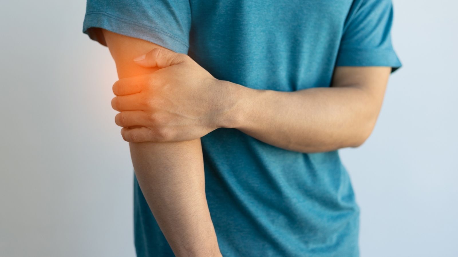
A stenosis or blockage in the iliac arteries often makes itself felt when you are on the move. When blood flow is reduced, especially during exertion, the muscles do not receive enough oxygen and this causes a range of symptoms. The most common symptom our patients present to us with is walking pain, also known as claudication, also known as “window disease”. This is a cramping, fatigue or tightness in the hip, thigh or calf muscles after walking a certain distance. The most typical feature of this pain is that it goes away completely when the person stops and rests for a few minutes. The location of the pain gives us an important clue about the level of congestion. Pain in the buttocks and thighs usually suggests that the problem is higher up, in the iliac arteries.
Symptoms that may occur as the disease progresses are as follows:
- Walking pain felt in the hip area
- Cramping or fatigue in the thigh muscles
- Pain in the calf (the blockage may also be further down)
- Difficulty walking at rest
- Erectile dysfunction, especially in male patients
- A constant feeling of chilliness or coldness in the legs and feet
- Pale or bruised skin
- Failure to heal even a simple injury to the feet or fingers
- Severe foot pain that starts at night when lying in bed and is relieved when the leg is lowered (rest pain)
- In the most advanced stage, blackening of the fingers (gangrene)
What is Leriche Syndrome and what are its symptoms?
Leriche Syndrome is an advanced and severe form of vascular occlusion that blocks the complete bifurcation of the aorta and the beginning of both main iliac arteries. In this case, blood cannot adequately reach both legs and the pelvic area. This syndrome manifests itself with a very typical triad of symptoms and the presence of this condition requires urgent and comprehensive treatment.
The classic symptom triad of Leriche Syndrome is as follows:
- Severe pain with walking in both hips and thighs
- Absence or very weak pulses in both groins on palpation
- Complete loss of erection in male patients
What is the Silent Danger of Iliac Artery Aneurysm?
In contrast to a blockage, an aneurysm is not a narrowing of the artery, but rather an abnormal weakening of the artery wall and its abnormal expansion, i.e. ballooning. An aneurysm of the iliac artery is when the wall of these vessels loses its elasticity and swells like a balloon. The most dangerous aspect of this disease is that it is often completely silent and asymptomatic. It is often detected by chance during an abdominal ultrasound or CT scan for another reason. The danger is that this balloon is subjected to constant pressure, grows larger over time and at some point bursts (rupture). Aneurysm rupture is a life-threatening emergency that causes uncontrolled internal bleeding into the abdomen and is characterized by sudden and severe pain.
Factors that increase the risk of developing an iliac artery aneurysm include
- over 60 years of age
- Male gender (more common than in women)
- Heavy smoking
- Uncontrolled high blood pressure
- Aneurysm disease in another family member
- Arteriosclerosis (atherosclerosis)
Which Diagnostic Methods are Used in Suspected Iliac Artery Disease?
When a patient complains of difficulty walking or leg pain, we follow a step-by-step diagnostic process to find the source of the problem. This process ranges from simple and painless tests to high-tech methods that map the vessels in detail when necessary. The aim is to make the correct diagnosis and create the most effective treatment plan.
The main methods we use in the diagnostic process usually follow the following sequence:
- Listening to the patient’s complaints in detail (patient history)
- Physical examination (especially checking pulses in the legs)
- Ankle-brachial pressure index (ABI) measurement
- Examination of blood flow with Color Doppler Ultrasonography
- Computed Tomography (CT) Angiography
- Magnetic Resonance (MR) Angiography
- Conventional Angiography (DSA – angiography) when necessary
How is the Ankle-Arm Index (ABI) Test Performed and What Does It Mean?
This test is a very simple screening method that gives us very fast and reliable information about the general condition of the blood flow in your leg veins. While you are lying on your back, we measure the blood pressure (systolic pressure) in both your arms and ankles using a sphygmomanometer and a handheld Doppler device. Then, for each leg, we divide the highest pressure measured at the ankle by the highest pressure measured at the arms. This ratio is an objective indicator of how much blood is flowing to your legs.
Interpretation of ABI test results is standardized
1.0 to 1.4: Normal blood flow
0.9 to 1.0: Considered borderline, requires follow-up
0.less than 9: Diagnosis of peripheral arterial disease (leg vascular occlusion)
0.7 to 0.9: Mild disease
0.4 to 0.7: Moderate disease
0.less than 4%: Severe disease and critical blood supply disturbance
What is the Basic Approach in the Treatment of Iliac Artery Stenosis?
Treatment of iliac artery disease is not just about opening a blocked vessel. It is the result of an underlying systemic problem of atherosclerosis. Therefore, no matter what treatment method is used, the basis of treatment is always lifestyle changes and medication. These steps are an absolute must to slow the progression of the disease, protect other vessels of the body (heart, brain) and ensure the longevity of treatment success.
These approaches form the basis of treatment:
- Strictly quit smoking and all tobacco products
- Regularly doing walking exercises every day, pushing to the limit of pain
- Switching to a Mediterranean-type diet low in salt and saturated fats and rich in vegetables and fruits
- Achieving and maintaining ideal weight
- Drugs that prevent blood clotting (such as Aspirin, Clopidogrel)
- Cholesterol-lowering drugs (Statins) are used to stabilize plaques even when cholesterol levels are normal
- Controlling high blood pressure with medication
- Strict control of blood glucose levels if diabetes is present
What is Closed Angioplasty and Stenting?
Today, minimally invasive, i.e. “closed” methods are the first choice for the treatment of stenosis and short obstructions in the iliac arteries. Angioplasty and stenting are the best known of these closed methods. The procedure is usually performed under local anesthesia by inserting a small needle through the artery in the groin. From this entry point, a thin wire is threaded through the vein and passed through the area of stenosis. A catheter with a small balloon at the tip is slid over the wire and inflated at the site of the stenosis to crush the plaque and widen the vessel. This procedure is called balloon angioplasty. To prevent the vessel from narrowing again and to provide it with a permanent support, a metal cage, called a stent, is often placed in the widened area.
This method has many advantages for patients:
- No large surgical incision
- Generally does not require general anesthesia
- Very short hospital stay (usually 1-2 days)
- Very little pain after the procedure
- Very quick return to daily life and work
- Lower risk of complications compared to open surgery
In Which Cases Is Open Surgery (Bypass Surgery) Necessary?
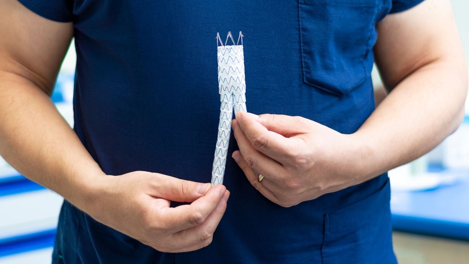
Open surgical methods still offer the most reliable and long-lasting solution in cases where closed methods are unsuitable due to the vessel structure or when the blockage is too long and complex. The most common open surgical procedure is aortobifemoral bypass surgery. The aim of this surgery is to create a new pathway for the blood to go around the blocked area. Through an incision in the abdomen, a “Y” shaped synthetic vessel (graft) is sewn to the intact part of the aorta. The two legs of this artificial vessel are connected to the intact femoral arteries below the blockage through small incisions in the groin. Thus, the blood reaches the legs via this new route, bypassing the blocked iliac arteries completely.
Situations where open surgery may be preferred are as follows:
- Very long and completely occluded vessel segments involving both iliac arteries
- Severe obstructions involving the bifurcation of the aorta (especially Leriche Syndrome)
- Cases where a previously tried closed method has failed or re-clogged
- Anatomical conditions where the vascular structure or calcification does not technically allow the closed method
- The search for the longest lasting and durable solution, especially in young and active patients
Which Treatment Method is Best for Me?
There is no single answer to this question. Because treatment is as individualized as a fingerprint. The right treatment method for you is determined by your age, your general health condition, the severity of your complaints and, most importantly, the structure of your vessels as revealed by angiographic images. Modern cardiovascular surgery offers us a wide range of treatments. For some patients, medication and lifestyle changes are sufficient, while for others a closed stenting procedure may be the best option. In some patients, the best and permanent solution is bypass surgery, which requires experience.
What is important here is that you are evaluated by an experienced team that is familiar with all these treatment options and a customized “road map” is drawn for you. The aim of the treatment is to relieve you from pain by restoring blood flow, to improve your quality of life and most importantly to prevent serious problems such as leg loss. Remember that with the right diagnosis and a tailor-made treatment, it is absolutely possible to overcome these problems and continue your life with healthy steps.

Prof. Dr. Yavuz Beşoğul graduated from Erciyes University Faculty of Medicine in 1989 and completed his specialization in Cardiovascular Surgery in 1996. Between 1997 and 2012, he served at Eskişehir Osmangazi University Faculty of Medicine as Assistant Professor, Associate Professor, and Professor, respectively. Prof. Dr. Beşoğul, one of the pioneers of minimally invasive cardiovascular surgery in Türkiye, has specialized in closed-heart surgeries, underarm heart valve surgery, beating-heart bypass, and peripheral vascular surgery. He worked at Florence Nightingale Kızıltoprak Hospital between 2012–2014, Medicana Çamlıca Hospital between 2014–2017, and İstinye University (Medical Park) Hospital between 2017–2023. With over 100 publications and one book chapter, Prof. Dr. Beşoğul has contributed significantly to the medical literature and is known for his minimally invasive approaches that prioritize patient safety and rapid recovery.